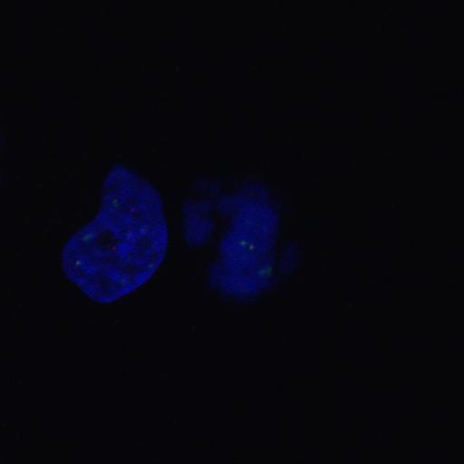
Hela cells were treated with anti-SENP6 siRNA and synchronized by double thymidine block. Metaphase cells were fixed and stained with CREST sera (red, Alexa Flour 568), CenpI antibody (green, Alexa Fluor 488) and Hoechst 33342 (blue). Fluorescence microscopy was performed at RT on a confocal microscope (LSM510 Meta; Carl Zeiss, Inc.) equipped with a 100× Plan-Apochromat objective. A 543 nm HeNe laser (5 mW output; detection LP560 nm) was used for detection of Alexa Fluor 568–labeled antibodies. The 488nm line of an Argon laser (25 mW nominal output; detection BP 505–530 nm) was used for analysis of Alexa Fluor 488–labeled antibodies. Hoechst 33258 images were captured using the 364nm line of an ion laser (Enterprise II ML UV; Coherent, Inc.; 80 mW nominal output; detection BP 385–470 nm). Image Reference: PMID 20212317
| Spatial Axis | Image Size | Pixel Size |
|---|---|---|
| X | 1024px | —— |
| Y | 1024px | —— |
| Z | 50px | —— |