Licensing
Description
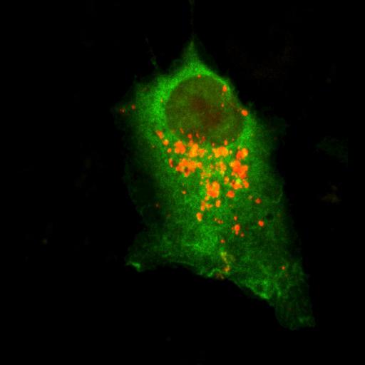
Image indicative of 24 h autophagic activity of human colon carcinoma HCT 116 cell co-transfected with a RFP-LC3-encoding plasmid and a SIRT1 variant with a mutation in the nuclear localization signal fused to GFP (mtNLS GFTP-SIRT1) which is virtually restricted to the cytoplasm. Confocal fluorescent images were captured using a confocal fluorescence microscope (TCS SP2; Leica) fitted with an Apochromat 63× 1.3 NA immersion objective. Images were acquired with a camera (DFC 350 FX 1.8.0; Leica) using LAS AF software (Leica) and processed with Photoshop (CS2; Adobe) software. Specifically, picture processing involved cropping of representative areas and linear adjustments of contrast and brightness and was performed using Photoshop (with equal adjustment parameters for all pictures); no explicit γ correction was used. Image: Figure 8C, bottom panel, in Morselli et al. J Cell Biol 192: 615-629
Biological Sources
- NCBI Organism Classification
- Homo sapiens
- (human)
- Cell Type
- permanent cell line cell
- Cell Line
- HCT 116
- Cellular Component
- autophagic vacuole
- cytoplasm
Biological Context
- Biological Process
- autophagy
- Molecular Function
- NAD-dependent protein deacetylase activity
Attribution
- Names
- Eugenia Morselli
- Guillermo Mariño
- Martin v. Bennetzen
- Tobias Eisenberg
- Evgenia Megalou
- Sabrina Schroeder
- Sandra Cabrera
- Paule Bénit
- Pierre Rustin
- Alfredo Criollo
- Oliver Kepp
- Lorenzo Galluzzi
- Shensi Shen
- Shoaib Ahmad Malik
- Maria Chiara Maiuri
- Yoshiyuki Horio
- Carlos López-Otín
- Jens S. Andersen
- Nektarios Tavernarakis
- Frank Madeo
- Guido Kroemer
- Published
- J Cell Biol 192: 615-629
- Link
- http://jcb-dataviewer.rupress.org/jcb/...
- Pubmed
- 21339330
Grouping
This image is part of a group.Imaging
- Image Type
- recorded image
- charge coupled device (CCD)
- Imaging Mode
- confocal microscopy
- Parameters Imaged
- fluorescence emission
- Source of Contrast
- distribution of a specific protein
- Visualization Methods
- EGFP
- Red fluorescent proteins from Anthozoa
- Processing History
- brightness and contrast adjusted
- Data Qualifiers
- processed data
Sample Preparation
- Methods
- formaldehyde fixed tissue
- Relation To Intact Cell
- whole mounted tissue
- cultured cells in vitro
Dimensions
| Spatial Axis | Image Size | Pixel Size |
|---|---|---|
| X | 1356px | —— |
| Y | 1492px | —— |