Alternate header for print version


Licensing
Attribution Non-Commercial Share Alike:This image is licensed under a Creative Commons Attribution, Non-Commercial Share Alike License. View License Deed | View Legal Code
tweet  
*CIL – Cell Image Library accession number. Please use this to reference an image.

CIL:13913*  Cite 
Description

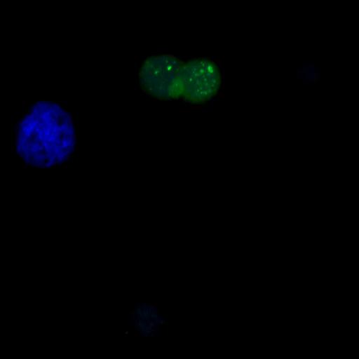
Human colon carcinoma HCT 116 cells were transfected with a GFP-LC3-encoding plasmid, cultured in complete medium for 24 h and enucleated to obtain cytoplasts. The cytoplasts were treated with 100 µM Spermidine for 4 h. Cytoplasts were still able to accumulate GFP-LC3 puncta in response to spermidine or resveratrol treatment, indicating that nuclei (and by extension transcription) are not required for short-term autophagy stimulation by these two agents. Nuclei were counterstained by Hoechst. Confocal fluorescent images were captured using a confocal fluorescence microscope (TCS SP2; Leica) fitted with an Apochromat 63× 1.3 NA immersion objective. Images were acquired with a camera (DFC 350 FX 1.8.0; Leica) using LAS AF software (Leica) and processed with Photoshop (CS2; Adobe) software. Specifically, picture processing involved cropping of representative areas and linear adjustments of contrast and brightness and was performed using Photoshop (with equal adjustment parameters for all pictures); no explicit γ correction was used. Image: Figure 8A, right panel, in Morselli et al. J Cell Biol 192: 615-629

Biological Sources
NCBI Organism Classification
Homo sapiens
Cell Type
permanent cell line cell
Cell Line
HCT 116
Cellular Component
autophagic vacuole
Biological Context
Biological Process
autophagy
Attribution
Names
Eugenia Morselli
Guillermo Mariño
Martin v. Bennetzen
Tobias Eisenberg
Evgenia Megalou
Sabrina Schroeder
Sandra Cabrera
Paule Bénit
Pierre Rustin
Alfredo Criollo
Oliver Kepp
Lorenzo Galluzzi
Shensi Shen
Shoaib Ahmad Malik
Maria Chiara Maiuri
Yoshiyuki Horio
Carlos López-Otín
Jens S. Andersen
Nektarios Tavernarakis
Frank Madeo
Guido Kroemer
Published
J Cell Biol 192: 615-629
Pubmed
21339330
Link
http://jcb-dataviewer.rupress.org/jcb/browse/...
Citation
Digital Object Identifier (DOI)
doi:10.7295/W9CIL13913
Archival Resource Key (ARK)
ark:/b7295/w9cil13913
Grouping This image is part of a group.
Imaging
Image Type
recorded image
charge coupled device (CCD)
Image Mode
confocal microscopy
Parameters Imaged
fluorescence emission
Source of Contrast
distribution of a specific protein
differences in adsorption or binding of stain
Visualization Methods
EGFP
membrane-permeant probe
Hoechst
Processing History
brightness and contrast adjusted
Data Qualifiers
processed data
Sample Preparation
Methods
isolated subcellular component
Relation To Intact Cell
enucleated cell
Dimensions
Spatial Axis Image Size Pixel Size
X 2048px ——
Y 2048px ——