Alternate header for print version


Licensing
Attribution Non-Commercial Share Alike:This image is licensed under a Creative Commons Attribution, Non-Commercial Share Alike License. View License Deed | View Legal Code
tweet  
*CIL – Cell Image Library accession number. Please use this to reference an image.

CIL:13902*  Cite 
Description

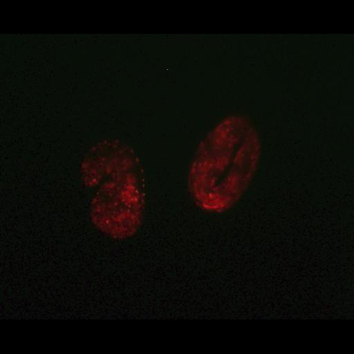
Representative image indicative of autophagic activity of C. elegans [VC199, sir2.1(ok434)IV] embryo expressing a full-length plgg-1DsRED::LGG-1 fusion fed with UV-killed Escherichia coli without additional 0.2 mM spermidine. Epifluorescence microscopy was performed with a microscope (AxioImager Z2; Carl Zeiss, Inc.) using a Plan Neofluar 40× objective with a 0.75 NA and a 63× Plan Neofluar objective with an NA of 1.25 in oil at RT using a 540 ± 15–nm band-pass excitation filter and a 575-nm long-pass emission filter. Images were taken with a camera (AxioCam MRc5; Carl Zeiss, Inc.) with Axiovision software (Carl Zeiss, Inc.) without further processing. Image: Figure 3A, bottom left panel, in Morselli et al. J Cell Biol 192: 615-629

Biological Sources
NCBI Organism Classification
Caenorhabditis elegans
Cell Type
embryo
Cell Line
VC199
Biological Context
Biological Process
autophagy
Attribution
Names
Eugenia Morselli
Guillermo Mariño
Martin v. Bennetzen
Tobias Eisenberg
Evgenia Megalou
Sabrina Schroeder
Sandra Cabrera
Paule Bénit
Pierre Rustin
Alfredo Criollo
Oliver Kepp
Lorenzo Galluzzi
Shensi Shen
Shoaib Ahmad Malik
Maria Chiara Maiuri
Yoshiyuki Horio
Carlos López-Otín
Jens S. Andersen
Nektarios Tavernarakis
Frank Madeo
Guido Kroemer
Published
J Cell Biol 192: 615-629
Pubmed
21339330
Link
http://jcb-dataviewer.rupress.org/jcb/browse/...
Citation
Digital Object Identifier (DOI)
doi:10.7295/W9CIL13902
Archival Resource Key (ARK)
ark:/b7295/w9cil13902
Grouping This image is part of a group.
Imaging
Image Type
recorded image
charge coupled device (CCD)
Image Mode
fluorescence microscopy
Parameters Imaged
fluorescence emission
Source of Contrast
distribution of a specific protein
Visualization Methods
DsRed
Data Qualifiers
raw, unprocessed data
Sample Preparation
Relation To Intact Cell
whole mounted tissue
Dimensions
Spatial Axis Image Size Pixel Size
X 1222px ——
Y 986px ——