Alternate header for print version


Licensing
Attribution Non-Commercial Share Alike:This image is licensed under a Creative Commons Attribution, Non-Commercial Share Alike License. View License Deed | View Legal Code
tweet  
*CIL – Cell Image Library accession number. Please use this to reference an image.

CIL:13899*  Cite 
Description

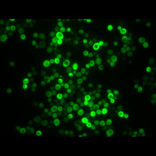
Saccharomyces cerevisiae (WT BY4741-MATa his3Δ1 leu2Δ0 met15Δ0 ura3Δ0) undergoing chronological aging on small synthetic 2% glucose media (SMD) with supplementation of 4 mM spermidine. Spermidine was added to stationary cultures at day 1 of the aging experiments. Image represents EGFP-Atg8p localization, visualized by fluorescence microscopy. Yeast cells undergoing autophagy exhibit a prominent vacuolar localization of EGFP-Atg8p. Epifluorescence image was captured using a widefield microscope (Axioskop; Carl Zeiss, Inc) fitted with a Plan Neofluar 63× 1.25 NA oil immersion objective. Image acquired with a camera (SPOT 9.0 Monochrome 6; Diagnostic Instruments, Inc) using Metamorph software (Universal Imaging Corp) and processed with IrfanView and Photoshop (CS2; Adobe) software. Specifically, picture processing involved coloring and cropping of representative areas and was performed with IrfanView. In addition, linear adjustments of contrast and brightness were applied with Photoshop (using equal adjustment parameters for all pictures); no explicit γ correction was used. Image: Figure 2A, WT/Spd/EGFP-Atg8p panel, in Morselli et al. J Cell Biol 192: 615-629

Biological Sources
NCBI Organism Classification
Saccharomyces cerevisiae S288c
Cell Line
BY 4741
Cellular Component
autophagic vacuole
Biological Context
Biological Process
autophagy
Attribution
Names
Eugenia Morselli
Guillermo Mariño
Martin v. Bennetzen
Tobias Eisenberg
Evgenia Megalou
Sabrina Schroeder
Sandra Cabrera
Paule Bénit
Pierre Rustin
Alfredo Criollo
Oliver Kepp
Lorenzo Galluzzi
Shensi Shen
Shoaib Ahmad Malik
Maria Chiara Maiuri
Yoshiyuki Horio
Carlos López-Otín
Jens S. Andersen
Nektarios Tavernarakis
Frank Madeo
Guido Kroemer
Published
J Cell Biol 192: 615-629
Pubmed
21339330
Link
http://jcb-dataviewer.rupress.org/jcb/browse/...
Citation
Digital Object Identifier (DOI)
doi:10.7295/W9CIL13899
Archival Resource Key (ARK)
ark:/b7295/w9cil13899
Grouping This image is part of a group.
Imaging
Image Type
recorded image
charge coupled device (CCD)
Image Mode
fluorescence microscopy
Parameters Imaged
fluorescence emission
Source of Contrast
distribution of a specific protein
Visualization Methods
EGFP
Processing History
brightness and contrast adjusted
Data Qualifiers
processed data
Sample Preparation
Relation To Intact Cell
whole mounted tissue
Dimensions
Spatial Axis Image Size Pixel Size
X 1360px ——
Y 1024px ——