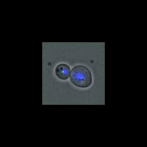
Tem1 normally localizes preferentially to the spindle pole body (SPB) that enters the daughter cell during anaphase (this image and CIL# 13882). This image shows SPB localization of eGFP-Tem1 (eGFP, green) in anaphase cells. tem1Δ::GAL-UPL-TEM1 cells expressing eGFP-TEM1 from a CEN plasmid were grown on 2% raffinose/2% galactose and transferred to 2% glucose medium prior to image capture. Nuclear morphology was assessed by DAPI (blue). A differential interference contrast (DIC) image is also shown (gray). The tem1Δ::GAL1-UPL-TEM1 strain allows for the rapid, conditional depletion of Tem1. UPL, which stands for ubiquitin-proline-LacI, acts as a destabilizing module that permits rapid degradation of appended proteins. Image is Fig 1B, top panels, in J Cell Biol. (2011) 192: 599-614. Other images in Fig 1 include CIL #13882, 13883, 13884, 13885, 13886, 13887.
Cells (MATa tem1::GAL-UPL-TEM1-TRP1 pRS316::eGFP-TEM1) were fixed in 2.5% formaldehyde for 10 min, washed twice, and resuspended in 0.1 M potassium phosphate buffer, pH 6.4. Cells were then fixed for 10 min in 80% ethanol and resuspended in 1 mg/ml DAPI. Imaging was performed at 25C using a Leica DM6000 microscope equipped with a 100x/1.40 NA oil immersion objective lens, A4, L5, and TX2 filters, and a digital CCD camera (DFC350, Leica). Pictures were processed with LAS AF (Leica) and ImageJ software.
| Spatial Axis | Image Size | Pixel Size |
|---|---|---|
| X | 218px | 0.0642µm |
| Y | 218px | 0.0642µm |