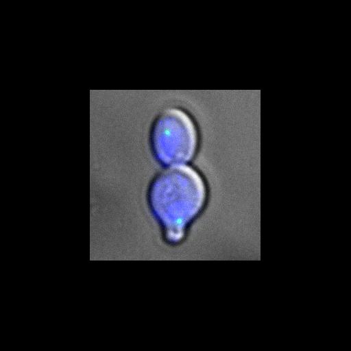
Tem1 (green) constitutively targeted to spindle pole bodies in nud1-2 cells at the restrictive temperature still displayed problems positioning the spindle (this image shows an aligned spindle, CIL #13889 shows a misaligned spindle and CIL# 13874 shows a rebudded cell) typical of nud1-2, despite the fact that viability was restored. Nud1 is required for mitotic exit and constitutive targeting of Tem1 recovered the mitotic exit defect of nud1-2 cells. DAPI (blue) and differential interference contrast (DIC) image are also shown. Image is Fig 7F, top panels, in J Cell Biol. (2011) 192: 599-614. Images in Fig 7 include CIL #13888, 13872, 13813, 13889, 13874.
Cells (MATa nud1::kanMX4 leu2::nud1-2::LEU2 pRS316::eGFP-CNM67–TEM1) grown at restrictive temperature 37C were fixed in 2.5% formaldehyde for 10 min, washed twice, and resuspended in 0.1 M potassium phosphate buffer, pH 6.4. Cells were then fixed for 10 min in 80% ethanol and resuspended in 1 mg/ml DAPI. Imaging was performed at 25C using a Leica DM6000 microscope equipped with a 100x/1.40 NA oil immersion objective lens, A4, L5, and TX2 filters, and a digital CCD camera (DFC350, Leica). Pictures were processed with LAS AF (Leica) and ImageJ software.
| Spatial Axis | Image Size | Pixel Size |
|---|---|---|
| X | 249px | 0.0642µm |
| Y | 249px | 0.0642µm |