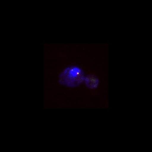
Both Tem1 (green) and Bfa1 (red) are localized to both spindle pole bodies in metaphase. DAPI (blue) and DIC (hidden) are also shown. Compare to anaphase image CIL# 13868. Image is Fig 6A, top panels, in J Cell Biol. (2011) 192: 599-614. Images in Fig 6 include CIL# 13869, 13868, 13871, 13870.
Cells (MATa tem1::TEM1-yEGFP::SpHIS5 bfa1::BFA1-mCherry-kanMX6) were fixed in 2.5% formaldehyde for 10 min, washed twice, and resuspended in 0.1 M potassium phosphate buffer, pH 6.4. Cells were then fixed for 10 min in 80% ethanol and resuspended in 1 mg/ml DAPI. Imaging was performed at 25C using a Leica DM6000 microscope equipped with a 100x/1.40 NA oil immersion objective lens, A4, L5, and TX2 filters, and a digital CCD camera (DFC350, Leica). Pictures were processed with LAS AF (Leica) and ImageJ software.
| Spatial Axis | Image Size | Pixel Size |
|---|---|---|
| X | 218px | 0.0642µm |
| Y | 218px | 0.0642µm |