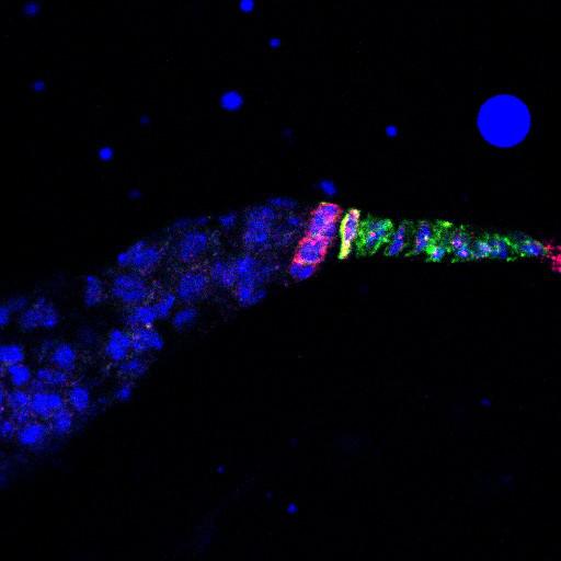
Boi (green) and Hedgehog (Hh, red) are expressed in apical cells of the Drosophila wild-type germarium (CIL# 13744). In the absence of boi (boi[e] mutant), Hh is redistributed from apical cells to the extracellular space of the local follicle stem cell niche (CIL# 13745). Expression of Boi in apical cells in the boi[e] mutant (boi[e]; UAS-Boi/+; babGal4/+) rescues Hh localization to apical cells (this image). However, expression of a Boi mutant lacking the Hh-binding domain does not (CIL# 13747). Nuclei are labeled (DRAQ5, blue). Image is Fig 3C in J Cell Biol. 2010. 191: 943-952. Images in Fig 3 include CIL# 13744, 13745, 13746, 13747, 13748.
Fly ovaries were dissected and fixed as described previously (O’Reilly et al., 2008). For anti-Boi immunostaining, ovaries were fixed in 2% formaldehyde on ice. For nuclear staining, fixed ovaries were incubated for 15 min with Draq5 (Cell Signaling Technology). Primary antibodies were 1:50 rat anti-Boi (Hartman et al., 2010); 1:100 goat anti-Hh (Santa Cruz Biotechnology, Inc.) or 1:100 rabbit anti-Hh (Ballet et al. 2003). Secondary antibodies used were FITC and Cy3 conjugated to species-specific secondary antibodies (Jackson ImmunoResearch Laboratories, Inc.). Samples were mounted in Vectashield mounting medium. Images were collected at room temperature (22C) using 40× (1.25 NA) or 63× (1.4 NA) oil immersion lenses (Leica) on an upright microscope (DM 5000; Leica) coupled to a confocal laser scanner (TCS SP5; Leica). LAS AF SP5 software (Leica) was used for data acquisition. Images representing individual channels of single confocal slices from the center of each germarium were exported as TIFF files, and images were converted to figures using Photoshop software (Adobe).
| Spatial Axis | Image Size | Pixel Size |
|---|---|---|
| X | 512px | 0.1991µm |
| Y | 512px | 0.1991µm |
| Z | 1px | 0.1998µm |