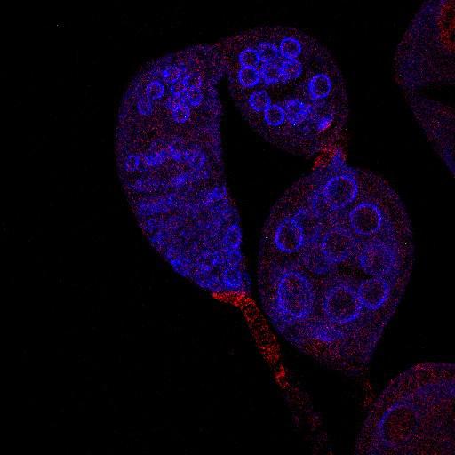
Boi (red) localizes predominantly to apical cells in Drosophila wild-type germaria but not in germaria with UAS-boi[RNAi] expression (CIL#13742). Germ cells are identified using anti-Vasa (blue). Image is Fig 2D in J Cell Biol. 2010. 191: 943-952. Other images in Fig 2 include CIL# 13738, 13739, 13740, 13741, 13742).
Fly ovaries were dissected and fixed as described previously (O’Reilly et al., 2008). Primary antibodies were 1:50 rat-antiBoi (Hartman et al. 2010) 1:2,000 rabbit anti-Vasa (Hay et al., 1990); Secondary antibodies used were Cy3 and Cy5 conjugated to species-specific secondary antibodies (Jackson ImmunoResearch Laboratories, Inc.). Samples were mounted in Vectashield mounting medium. Images were collected at room temperature (22C) using 40× (1.25 NA) or 63× (1.4 NA) oil immersion lenses (Leica) on an upright microscope (DM 5000; Leica) coupled to a confocal laser scanner (TCS SP5; Leica). LAS AF SP5 software (Leica) was used for data acquisition. Images representing individual channels of single confocal slices from the center of each germarium were exported as TIFF files, and images were converted to figures using Photoshop software (Adobe).
| Spatial Axis | Image Size | Pixel Size |
|---|---|---|
| X | 512px | 0.2342µm |
| Y | 512px | 0.2342µm |
| Z | 1px | 0.042µm |