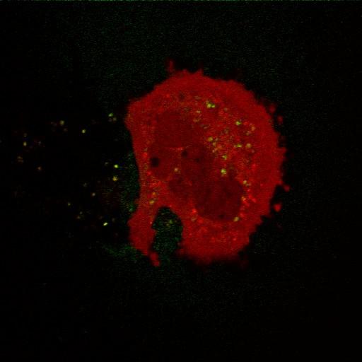
Expression of WT PINK1-YFP (green) does not induce mitochondrial translocation of mCherry-Parkin (red) in PINK1 knock-out mouse embryonic fibroblasts (PINK1 KO MEFs). Transfected PINK1 KO MEFs were treated with DMSO for 3 hrs. Live cell imaging was performed on an LSM510 Meta (Carl Zeiss, Inc) with a 63x 1.4 NA oil differential interference contrast Plan Apo objective. Image contrast and brightness were adjusted in the LSM image browser (Zeiss). This image corresponds to Supplemental Figure S1c, top left row of J Cell Biol, 191: 933-942, 2010. Images in Supplemental Figure S1c include CIL#s 13725, 13726, 13727, 13728.
| Spatial Axis | Image Size | Pixel Size |
|---|---|---|
| X | 512px | 0.1395µm |
| Y | 512px | 0.1395µm |