Licensing
Description
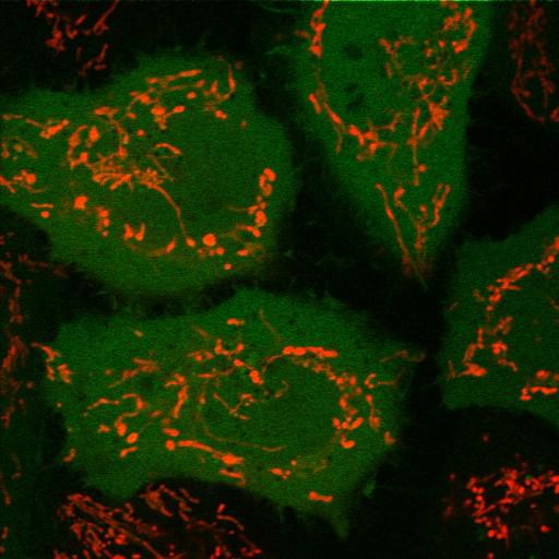
YFP-Parkin (green) does not translocate to mitochondria (red) when HeLa cells are transfected with PARL (presenilin-associated rhomboid-like protein) siRNA. HeLa cells stably expressing YFP-Parkin were transfected with PARL siRNA for 192 hours, treated with DMSO for 1 hour and then stained with TMRE, a cationic fluorescent dye that is readily sequestered by active mitochondria. Cells were imaged using an inverted microscope (LSM510 Meta; Carl Zeiss, Inc.) with a 63× 1.4 NA oil differential interference contrast Plan Apo objective. Image contrast and brightness were adjusted in the accompanying image browser (LSM; Carl Zeiss, Inc.). Image corresponds to Fig1f, bottom panels in J Cell Biol. 191: 933-942, 2010. Images in Fig1f include CIL#s 13707, 13708, 13709.
Biological Sources
- NCBI Organism Classification
- Homo sapiens
- (human)
- Cell Type
- epithelial cell
- cervical carcinoma
- Cell Line
- HeLa
- Cellular Component
- mitochondrion
- cytosol
- nucleus
- mitochondrial inner membrane
Biological Context
- Biological Process
- mitochondrion degradation
- protein K48-linked ubiquitination
- protein K63-linked ubiquitination
- protein ubiquitination involved in ubiquitin-dependent protein catabolic process
- Molecular Function
- ubiquitin-protein ligase activity
- zinc ion binding
- protein binding
- serine-type endopeptidase activity
Attribution
- Names
- Seok Min Jin
- Michael Lazarou
- Chunxin Wang
- Lesley A. Kane
- Derek P. Narendra
- Richard J. Youle
- Published
- J Cell Biol. 191: 933-942, 2010.
- Link
- http://jcb-dataviewer.rupress.org/jcb/...
- Pubmed
- 21115803
Grouping
This image is part of a group.Imaging
- Image Type
- recorded image
- Imaging Mode
- single-spot confocal microscopy
- Parameters Imaged
- fluorescence emission
- Source of Contrast
- distribution of a specific protein
- compartmentalization of stain or label
- Visualization Methods
- EYFP
- probe for mitochondria
- tetramethylrhodamine, ethyl ester (TMRE)
- Processing History
- brightness and contrast adjusted
- Data Qualifiers
- raw, unprocessed data
- suitable for spatial measurements
Sample Preparation
- Methods
- living tissue
- Relation To Intact Cell
- dispersed cells in vitro
Dimensions
| Spatial Axis | Image Size | Pixel Size |
|---|---|---|
| X | 512px | .1395μm |
| Y | 512px | .1395μm |