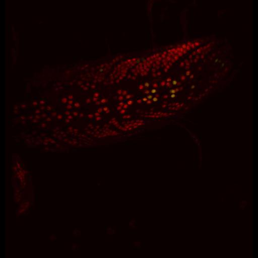
The JNK phosphatase puckered, whose expression can be reported via a lacZ enhancer trap, is expressed in very low levels in uninjured neurons; however, nerve crush induces a dramatic increase in puc-lacZ expression in injured motoneurons. This image stack shows puc-lacZ (green) and neuronal nuclei (red) in a third instar larva injred at site 3 (as defined in Fig 1C). Wandering third instar larvae were dissected in PBS and fixed in either 4% paraformaldehyde in PBS or Bouin’s fixative for 15–30 min. Primary antibodies used were: rat anti-elaV (7E8A10, Developmental Studies Hybridoma Bank) and mouse anti-lacZ (40-1a, DSHB). Secondary antibodies were Alexa 488 anti-mouse and Cy3 anti-rat. Confocal images were collected at room temperature on a spinning-disk confocal system (PerkinElmer) consisting of a scanner (Nipkow CSU10; Yokogawa) and an electron microscopy charge-coupled device camera (C9100-50; Hamamatsu Photonics) mounted on an inverted microscope (Axio Observer; Carl Zeiss, Inc.) with 25× 0.8 NA multi and 40× 1.3 NA, 63× 1.5 NA, and 100× 1.46 NA oil objectives. Similar settings were used to collect all compared genotypes. All imaging and quantification were conducted with Volocity software (PerkinElmer). This image stack corresponds to Fig 1E3 in J Cell Biol. 2010. 191: 211-223. Images in Fig 1 include CIL#s 13676, 13677, 13678, 13679.
| Spatial Axis | Image Size | Pixel Size |
|---|---|---|
| X | 962px | 0.5869µm |
| Y | 1000px | 0.5869µm |
| Z | 8px | 0.8µm |