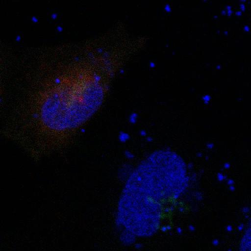
A major proportion of the medial-golgi enzyme tagged with HA, GlcNAcT-1-HA (green), is mislocalized to the ER when coexpressed with Myc-GnT1IP-L (Myc-tagged GlcNAcT-1 inhibitory protein, long transcript, membrane-anchored) (red). Transiently expressed Myc-GnT1IP-L itself is normally primarily localized in the Golgi and ERGIC and partially in the ER (shown in Fig 4; J Cell Biol. 190: 893-910, 2010.). Transfected HeLa cells were fixed in 3% paraformaldehyde, blocked with 0.2% Triton X-100, 1% FBS, 0.5% BSA in PBS with Ca[2+] and Mg[2+], followed by incubation in primary antibodies (anti-Myc mouse mAB, 9E10 (Covance) and anti-HA mouse mAb, HA.11 (Covance) and secondary antibodies (Alexa 568 and Alexa 488) and DAPI (blue) to stain nuclei. Cells were mounted using Fluoromount (SouthernBiotech). Images of confocal microscopy were acquired by capturing Z-series images with a 0.25-µm step size on a confocal microscope (TCS SP2 AOBS; Leica) using a 63x/1.4 NA oil immersion objective (HCX PL APO λBL CS; Leica). Laser lines at 405-, 488-, and 561-nm were provided by 20 mW diode, 100 mW Ar, and 10 mW diode, respectively; sequential excitation by line and detection range settings was used to eliminate cross talk between fluorophores. The images (512 × 512 pixel, 8 bit) were saved as tiff files. The entire Z-series was projected using the sum intensity method provided by ImageJ (NIH). This image corresponds to Figure 5A, top panel (merge) in J Cell Biol. 190: 893-910, 2010. Images in Fig 5 include CIL#s 13658, 24919, 24920.
| Spatial Axis | Image Size | Pixel Size |
|---|---|---|
| X | 512px | 0.465µm |
| Y | 512px | 0.465µm |
| Z | 17px | 0.0407µm |