Alternate header for print version


Licensing
Attribution Non-Commercial Share Alike:This image is licensed under a Creative Commons Attribution, Non-Commercial Share Alike License. View License Deed | View Legal Code
tweet  
*CIL – Cell Image Library accession number. Please use this to reference an image.

CIL:13654*  Cite 
Description

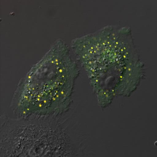
Image is Fig. 4D in PMID: 20679433. HeLa cells were transfected to co-express p125A-mCherry and Sec31A-GFP. p125A colocalizes extensively with Sec31A; live cell time-lapse video microscopy showed that p125A (red) and Sec31A (green) positive structures are colocalized stably with each other over time, and undergo homotypic fusion. The vesicular structures of p125A-mCherry colocalize extensively with those of Sec31A-GFP over a period of at least 30 min. Cells were cultured on glass-bottom culture dishes, transfected with plasmid constructs of interest, and imaged using the Olympus Fluoview 1000 confocal microscope. 60X objective.

Biological Sources
NCBI Organism Classification
Homo sapiens
Cell Line
HeLa
Cellular Component
trans-Golgi network
Biological Context
Biological Process
Golgi organization
Attribution
Names
Yan Shan Ong
Bor Luen Tang
Li ShenLoo
Wanjin Hong
Pubmed
PMID:20679433
Citation
Digital Object Identifier (DOI)
doi:10.7295/W9CIL13654
Archival Resource Key (ARK)
ark:/b7295/w9cil13654
Grouping This image is part of a group.
Sample Preparation
Methods
formaldehyde fixed tissue
Relation To Intact Cell
whole mounted tissue
Dimensions
Spatial Axis Image Size Pixel Size
X 512px ——
Y 512px ——