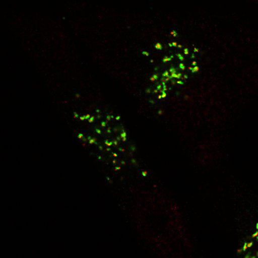
This image is Fig. 6A (p125A siRNA). HeLa cells stably expressing ManII-GFP were silenced of p125A and then double labeled using mouse anti-GM130 (secondary Ab: goat anti-mouse Ab labeled with Alexa 633, shown in blue) and rabbit anti-GT (secondary Ab: goat anti-rabbit Ab labeld with Alex 555, shown in red) antibodies followed by viewing the distribution of trans-Golgi marked by GT, medial-Golgi marked by ManII-GFP (shown in green), and cis-Golgi marked by GM130. Technical details: Cells grown on coverslips were washed 2X with PBS supplemented with 1 mM CaCl2 and 1 mM MgCl2 (PBSCM), fixed with 4% paraformaldehyde in PBSCM for 20 min at RT, washed 5X at 5-min intervals using PBSCM, and then permeabilized with 0.1% Saponin in PBSCM for 20 min at RT. The cells were then immunolabeled with appropriate primary antibodies diluted in fluorescence dilution buffer (FDB; PBSCM with 5% FBS and 2% bovine serum albumin [BSA]) for 1 h at RT, washed 5X with 0.1% Saponin PBSCM at 5-min intervals. Secondary antibodies were diluted in FDB and incubated at RT for 1 h, washed again with 0.1% Saponin PBSCM 5X at 5-min intervals, and then 2X with PBSCM. The coverslips were mounted on microscopic slides with Vectashield mounting medium containing DAPI. Confocal microscopy was performed with an Axioplan II microscope (Carl Zeiss, Inc.) equipped with Zeiss confocal scanning optics. 40x objective.
| Spatial Axis | Image Size | Pixel Size |
|---|---|---|
| X | 512px | —— |
| Y | 512px | —— |
| Z | 5px | —— |