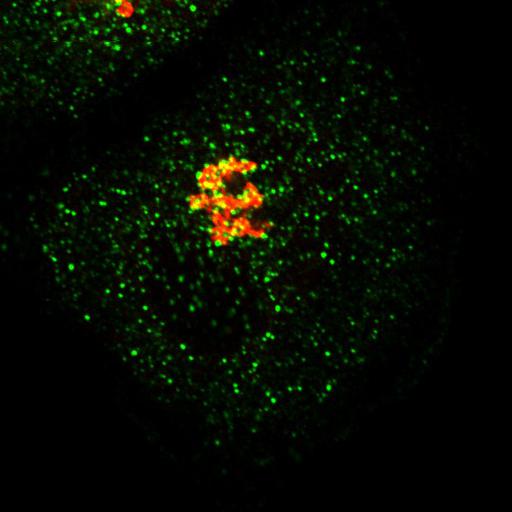
This image is Fig. 4B from PMID: 20679433. HeLa cells were fixed and double-labeled with rabbit anti-p125A (secondary Ab: goat anti-rabbit Ab conjugated with Alexa 555, shown in red) and mouse anti-Sec31A (secondary Ab: goat anti-mouse Ab conjugated with FITC, shown in green) antibodies. Technical details: Cells grown on coverslips were washed 2X with PBS supplemented with 1 mM CaCl2 and 1 mM MgCl2 (PBSCM), fixed with 4% paraformaldehyde in PBSCM for 20 min at RT, washed 5X at 5-min intervals using PBSCM, and then permeabilized with 0.1% Saponin in PBSCM for 20 min at RT. The cells were then immunolabeled with appropriate primary antibodies diluted in fluorescence dilution buffer (FDB; PBSCM with 5% FBS and 2% bovine serum albumin [BSA]) for 1 h at RT, washed 5X with 0.1% Saponin PBSCM at 5-min intervals. Secondary antibodies were diluted in FDB and incubated at RT for 1 h, washed again with 0.1% Saponin PBSCM 5X at 5-min intervals, and then 2X with PBSCM. The coverslips were mounted on microscopic slides with Vectashield mounting medium containing DAPI. Confocal microscopy was performed with an Axioplan II microscope (Carl Zeiss, Inc.) equipped with Zeiss confocal scanning optics. 100x objective.
| Spatial Axis | Image Size | Pixel Size |
|---|---|---|
| X | 512px | —— |
| Y | 512px | —— |
| Z | 5px | 0.5µm |