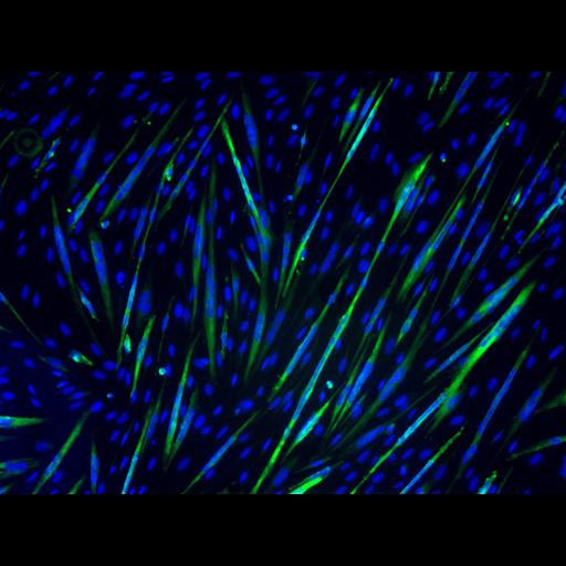
The C2C12 cell line, a mouse myoblast line, was used here to study the regulatory factors in myogenic differentiation. After cultured in differentiation medium for 3 days, these cells differentiated into myotubes (Green) containing multiple nuclei (Blue). The C2C12 cells stably expressing FLAG-tagged rapamycin-resistant, kinase-inactive (RR/KI) mTOR were induced to differentiate in the presence of 50nM rapamycin (Rap) for 3 days. Conditioned medium (CM) was collected daily from parental C2C12 cells that had been induced to differentiate 1 day earlier than the RR/KI mTOR cells and fed to the latter with 50nM Rap added. CM was able to rescue the effect of Rap to allow myotube maturation. This image is the control of different treatments in Figure 9B from JCB 189: 1157-1169, 2010. See also CIL: 13592, 13593, 13601.
The image was taken by leica DMI 4000B microscope equipped with a QImaging RETIGA Exi camera and Leica 10X/0.22 lens. Myosin heavy chain (MHC) expressed in myotubes was labeled by MF-20 mouse antibody followed by FITC-anti-mouse IgG secondary antibody. Nuclei were stained by DAPI. MHC and nuclei were pseudocolored in green and blue, respectively.
| Spatial Axis | Image Size | Pixel Size |
|---|---|---|
| X | 1392px | 0.645µm |
| Y | 1040px | 0.645µm |