Licensing
Description
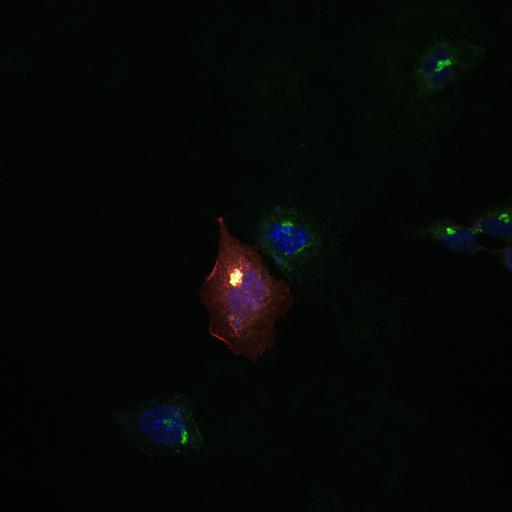
Co-expression of wild-type Arf1-GFP (Arf1(WT)-GFP) (gray) and inactive Src (SrcK-DsRed) (red) in Src-deficient SYF fibroblasts does not induce trafficking of Helix Pomatia Lectin (HPL) (green) from the Golgi. HPL binds various glycans but the Tn antigen in particular. The Tn antigen refers to terminal α-linked N-acetyl galactosamine residues (GalNAc) linked to Ser or Thr residues. HPL indicates distribution of GalNac-Ts. Inactive Src is highly enriched at the Golgi. Cells were fixed for 10 min (4% paraformaldehyde) and permeabilized (0.2% Triton X-100) prior to staining with Hoechst (blue) and Alexa 647-conjugated-HPL. Cells were mounted onto glass slides using FluorSave (Merck) and imaged at room temperature using an inverted FluoView confocal microscope (model IX81; Olympus) with either a 60x objective (U Plan Super Apochromatic, UPLSAPO; NA 1.35) or 100x objective (UPLSAPO; NA 1.40) using Immersol oil. Microscope coupled with a CCD camera (model FVII). Images were acquired and processed using Olympus FV10-ASW software. Image corresponds to Fig 7F in J Cell Biol. 189: 843-858. 2010. Images in Fig 7 include CIL#s 13571, 13572, 13573, 13574, 13575, 13576, 13577.
Biological Sources
- NCBI Organism Classification
- Homo sapiens
- (human)
- Cell Type
- epithelial cell
- cervical carcinoma
- Cell Line
- HeLa
- Cellular Component
- Golgi stack
- integral to membrane
- nucleus
Biological Context
- Biological Process
- Golgi organization
- COPI coating of Golgi vesicle
- retrograde vesicle-mediated transport, Golgi to ER
- small GTPase mediated signal transduction
- Molecular Function
- GTPase activity
- protein binding
Attribution
- Names
- David J. Gill
- Joanne Chia
- Jamie Senewiratne
- Frederic Bard
- Published
- J Cell Biol. 189: 843-858. 2010.
- Link
- http://jcb-dataviewer.rupress.org/jcb/...
- Pubmed
- 20498016
Grouping
This image is part of a group.Imaging
- Image Type
- recorded image
- Imaging Mode
- single-spot confocal microscopy
- Parameters Imaged
- fluorescence microscopy
- Source of Contrast
- distribution of a specific protein
- distribution of epitope
- compartmentalization of stain or label
- Visualization Methods
- Alexa Fluor 647
- Alexa Fluor 594
- EGFP
- primary antibody plus labeled secondary antibody
- Helix pomatia agglutinin
- Hoechst
- Data Qualifiers
- raw, unprocessed data
- suitable for spatial measurements
Sample Preparation
- Methods
- formaldehyde fixed tissue
- detergent permeabilized
- Relation To Intact Cell
- dispersed cells in vitro
Dimensions
| Spatial Axis | Image Size | Pixel Size |
|---|---|---|
| X | 1024px | .2070μm |
| Y | 1024px | .2070μm |
| Z | 1 | 13.9500μm |