Alternate header for print version


Licensing
Attribution Non-Commercial Share Alike:This image is licensed under a Creative Commons Attribution, Non-Commercial Share Alike License. View License Deed | View Legal Code
tweet  
*CIL – Cell Image Library accession number. Please use this to reference an image.

CIL:13560*  Cite 
Description

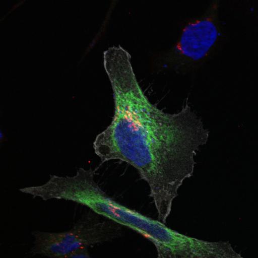
An ER-trapped GalNac-T activity reporter (Muc-PTS; fused to GFP) (green) is targeted to the ER and not the Golgi, marked by Giantin (red) in HeLa cells cotransfected with inactive Src (SrcK-mCherry, containing K295M mutation) (gray). Cells were fixed for 10 min (4% paraformaldehyde) and permeabilized (0.2% Triton X-100). Primary antibody staining followed the manufacturer’s instructions. Cells were subsequently stained for 15–30 min with secondary Alexa 647 for anti-Giantin and Hoechst (blue). Cells were mounted onto glass slides using FluorSave (Merck) and imaged at room temperature using an inverted FluoView confocal microscope (model IX81; Olympus) with fluorescence excitation at 488 nm, 561 nm and 633 nm and either a 60x objective (U Plan Super Apochromatic [UPLSAPO]; NA 1.35) or 100x objective (UPLSAPO; NA 1.40) using Immersol oil. Microscope coupled with a CCD camera (model FVII). Images were acquired and processed using Olympus FV10-ASW software. Image corresponds to Fig 5C in J Cell Biol. 189: 843-858. 2010. Images in Fig 5 include CIL#s 13559, 13560, 13561, 13562.

Biological Sources
NCBI Organism Classification
Homo sapiens
Cell Type
epithelial cell
cervical carcinoma
Cell Line
HeLa
Cellular Component
Golgi stack
integral to membrane
endoplasmic reticulum
nucleus
Attribution
Names
David J. Gill
Joanne Chia
Jamie Senewiratne
Frederic Bard
Published
J Cell Biol. 189: 843-858. 2010.
Pubmed
PMID: 20498016
Link
http://jcb-dataviewer.rupress.org/jcb/browse/...
Citation
Digital Object Identifier (DOI)
doi:10.7295/W9CIL13560
Archival Resource Key (ARK)
ark:/b7295/w9cil13560
Grouping This image is part of a group.
Imaging
Image Type
recorded image
Image Mode
single-spot confocal microscopy
Parameters Imaged
fluorescence microscopy
Source of Contrast
distribution of a specific protein
compartmentalization of stain or label
Visualization Methods
Alexa Fluor 647
EGFP
mCherryFP
primary antibody plus labeled secondary antibody
Hoechst
Data Qualifiers
raw, unprocessed data
suitable for spatial measurements
Sample Preparation
Methods
formaldehyde fixed tissue
detergent permeabilized
Relation To Intact Cell
dispersed cells in vitro
Dimensions
Spatial Axis Image Size Pixel Size
X 1024px 0.103µm
Y 1024px 0.103µm
Z 1px 3.09µm