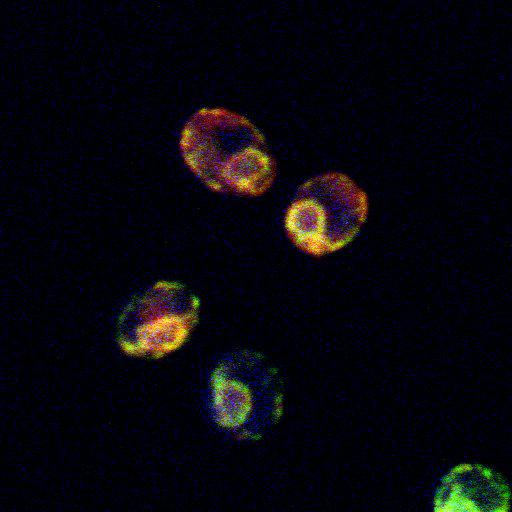
PrA contains a distinct determinant for glycan-independent ERAD (ER associated degradation). A mutant of the vacuolar proteinase A (PrA*-Gldelta4) (green) is a substrate of ER quality control (ERQC) mechanisms, as determined by the fact that it is retained in the ER (marked by Kar2; red), but is incompetent for ERAD. S. cerevisiae wild-type bearing PrA*-Gldelt4-HA centromeric plasmid were fixed in 3.7% formaldehyde. The cell wall was removed by zymolyase digestion in 1.2M sorbitol and washed resuspended cells were applied to poly L-lysine coated slide, methanol (-20C) and acetone treated, and air dried. Primary antibodies HA.11 mAb (Covance) and polyclonal rabbit anti-Kar2p were applied in 0.05% Tween 20 followed by Alexa Fluor 488 goat anti–mouse and Alexa Fluor 594 goat anti–rabbit (Invitrogen) secondary antibodies. Slides were mounted in PBS, 90% glycerol and DAPI (blue). Cells were visualized using an inverted microscope (LSM 510 META; Carl Zeiss, Inc.) with a Plan Apochromat 100× 1.4 NA Ph3 objective (Carl Zeiss, Inc.) in immersion oil (Immersol 518F; Carl Zeiss, Inc.) at room temperature. Image acquisition was performed using standard photomultiplier tube with LSM 510. Images were archived using LSM 5 Image Examiner (Carl Zeiss, Inc.) and Photoshop (version 7.0; Adobe), and no additional software adjustments were performed on images after acquisition other than cropping. Image corresponds to Fig 5D in J Cell Biol. 2010. 188:707-716.
| Spatial Axis | Image Size | Pixel Size |
|---|---|---|
| X | 512px | 0.0464µm |
| Y | 512px | 0.0464µm |
| Z | 1px | 0.5453µm |