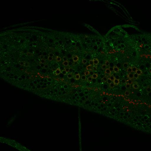
Colocalization between DVGLUT (red) aggregates and the endosomal ESCRT protein Hrs (green, also known as Vps27) in motoneuron cell bodies of the ventral nerve cord of a wild-type larva. Wandering third instar larvae were dissected in ice-cold PBS and fixed with Bouin’s solution for 5 min. Blocking and antibody incubation were performed in PBS containing 0.1% Triton X-100. anti-DVGLUT (Daniels et al., 2004) antibody was used at 1: anti-HRS-FL (Lloyd et al., 2002) was used at 1:1000. Secondary antibodies (Alexa 488-/Cy3-conjugated) were used at 1:1000. After staining, specimens were equilibrated in 70% glycerol in PBS and mounted with VectaShield (Vector Laboratories). Confocal images were acquired with a confocal microscope (model C1; Nikon) and accompanying EZ-C1 software using argon (excitation at 488 nm) and HeNe (excitation at 543 and 633 nm) lasers and a 60x Plan-Apochromat NA 1.4 objective (Nikon) at room temperature. Samples for each experiment were processed using the same confocal gain setting. Image corresponds to Figure 2A, top 3 panels in Kim et al. J Cell Biol. 188: 717-734. 2010.
| Spatial Axis | Image Size | Pixel Size |
|---|---|---|
| X | 2048px | —— |
| Y | 2048px | —— |