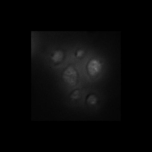
Intracellular localization of GFP-Vps74 in a wild-type S. cerevisiae background grown at 37C. In wild-type cells incubated at 26C or 37C, GFP-Vps74 is localized to Golgi compartments and the cytosol. The Golgi-localized PtdIns 4-kinase in yeast is composed of the Pik1 catalytic subunit and a myristoylated calcium-binding protein, Frq1. Vps74 is essential to maintain glycosyltransferases in the Golgi and results in this study reveal a previously unrecognized PtdIns4P-binding site in Vps74/GOLPH3 family proteins and link Pik1 signaling to retention of Golgi-resident proteins. Cells grown in liquid medium were mounted in growth medium and 3D image stacks were collected at 0.4-µm z increments on a DeltaVision workstation (Applied Precision) based on an inverted microscope (IX-70; Olympus) using a 100× NA 1.4 oil immersion lens. Images were captured at 23C with a 12-bit CCD camera (CoolSnap HQ; Photometrics) and deconvolved using the iterative-constrained algorithm (Agard, 1984) and the measured point spread function. One image from the approximate center of z stack is shown in Fig1A Vps74/wild-type 37C panel in J Cell Biol. 187: 967-975. 2009. Images in Fig 1A include CIL#s 13439, 13440, 13441, 13442, 13443, 13444, 13445, 13446, 13447, 13448, 13458, 13459.
| Spatial Axis | Image Size | Pixel Size |
|---|---|---|
| X | 302px | 0.0663µm |
| Y | 302px | 0.0663µm |
| Z | 12px | 0.4µm |