Alternate header for print version


Licensing
Attribution Non-Commercial Share Alike:This image is licensed under a Creative Commons Attribution, Non-Commercial Share Alike License. View License Deed | View Legal Code
tweet  
*CIL – Cell Image Library accession number. Please use this to reference an image.

CIL:13384*  Cite 
Description

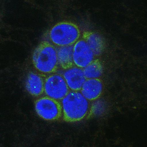
FISH detection of PTEN and VEGF gene loci in MCF-10.B2 cells during mammary epithelial differentiation and early mammary tumorigenesis. PTEN (red) and VEGF (green)were detected in paraformaldehyde fixed MCF10A.B2 cells grown for 20 days under 3D growth conditions. DAPI (blue) delineates nuclei. A representative acinus structure is shown. The z-series was recorded with a Zeiss LSM 510 META confocal microscope with a 63x 1.4 NA objective lens and zoom level 3. Optical sections imaged totaled approximately 15-20 microns in thickness. See Fig 2 in Meaburn and Misteli, 2008 J Cell Biol 180:39-50.

Biological Sources
NCBI Organism Classification
Homo sapiens
Cell Type
mammary epithelia (MEC)
Cell Line
MCF-10A.B2
Cellular Component
nuclear chromatin
Biological Context
Biological Process
tumorigenesis
Attribution
Names
Karen Meaburn
Tom Misteli
Published
Meaburn KJ, Misteli T 2008 J Cell Biol 180:39-50
Pubmed
18195100
Citation
Digital Object Identifier (DOI)
doi:10.7295/W9CIL13384
Archival Resource Key (ARK)
ark:/b7295/w9cil13384
Grouping This image is part of a group.
Sample Preparation
Methods
formaldehyde fixed tissue
Relation To Intact Cell
whole mounted tissue
Dimensions
Spatial Axis Image Size Pixel Size
X 1024px ——
Y 1024px ——
Z 53px 0.3µm