Alternate header for print version


Licensing
Attribution Non-Commercial Share Alike:This image is licensed under a Creative Commons Attribution, Non-Commercial Share Alike License. View License Deed | View Legal Code
tweet  
*CIL – Cell Image Library accession number. Please use this to reference an image.

CIL:13382*  Cite 
Description

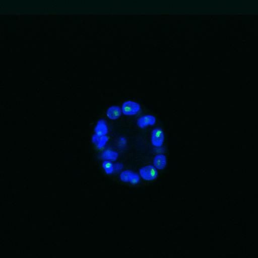
The nucleolus during mammary epithelial differentiation and early mammary tumorigenesis. Proliferating 3D cultures of MCF-10.B2 cells at day 20 were stained with ANA-N antibody to detect nucleoli (green) and counterstained with DAPI to delineate nuclei (blue). Under these conditions, cells form acini. The stack shown is presented as a projected image in Fig 1A (Day 20 control)of Meaburn and Misteli, 2008 J Cell Biol 180:39-50. The z-series was recorded with a Zeiss LSM 510 META confocal microscope with a 63x 1.4 NA objective lens.

Biological Sources
NCBI Organism Classification
Homo sapiens
Cell Type
mammary epithelia (MEC)
Cell Line
MCF-10A.B2
Cellular Component
nuclear chromatin
nucleolus
Biological Context
Biological Process
tumorigenesis
Attribution
Names
Karen Meaburn
Tom Misteli
Published
Meaburn KJ, Misteli T 2008 J Cell Biol 180:39-50
Pubmed
18195100
Citation
Digital Object Identifier (DOI)
doi:10.7295/W9CIL13382
Archival Resource Key (ARK)
ark:/b7295/w9cil13382
Grouping This image is part of a group.
Imaging
Image Type
recorded image
Image Mode
confocal microscopy
Parameters Imaged
fluorescence emission
Source of Contrast
differences in adsorption or binding of stain
Visualization Methods
Alexa Fluor 488
4',6-diamidino-2-phenylindole (DAPI)
Processing History
unprocessed raw data
Sample Preparation
Methods
formaldehyde fixed tissue
Relation To Intact Cell
whole mounted tissue
Dimensions
Spatial Axis Image Size Pixel Size
X 1024px ——
Y 1024px ——
Z 21px 0.3µm