Licensing
Attribution Non-Commercial Share Alike:This image is licensed under a Creative Commons Attribution, Non-Commercial Share Alike License. View License Deed | View Legal Code
Comments:
Powered by Pnyxe
Description
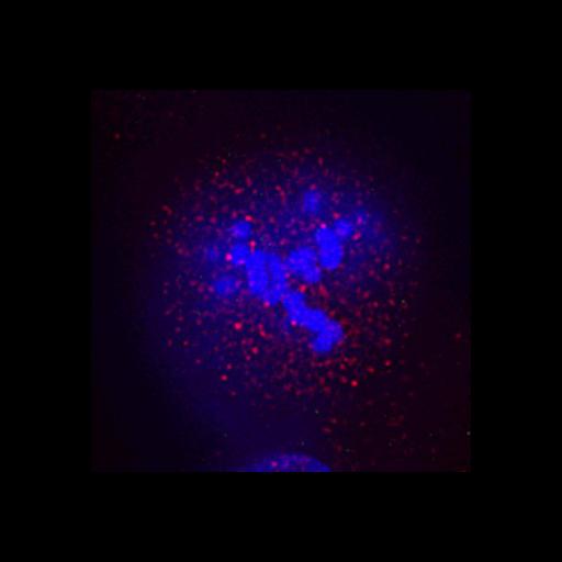
Z-focal series of HeLa cells at metaphase. Cells were stained for DNA (blue), phosphorylated mitotic centromere-associated kinesin (MCAK) (red) and HEC1 (green). Cells were transfected with control (this image) or Bod1 (CIL:13379) siRNA. After 72 h, cells were treated with monastrol for 3 h and released into media containing MG132 for 1 h before fixing.
Technical Details
Antibodies were rabbit anti-HEC1 (green) antibody (Abcam) used at 1:1,000. and anti–phospho-MCAK antibodies (red) (Andrews et al., 2004) used at 1 μg/ml, and DNA (blue). Fluorescently labeled secondary antibodies were all obtained from Jackson ImmunoResearch Laboratories. Corresponds to Fig 5B in JCB vol. 179 no. 2 187-197.
Biological Sources
- NCBI Organism Classification
- Homo sapiens
- (human)
- Cell Line
- HeLa S3
- Cellular Component
- chromosome, centromeric region
- DNA
- phosphorylated MCAK
- HEC1
- nuclear chromosome
Biological Context
- Biological Process
- mitosis
- Bod1 SiRNA
- chromosome organization
Attribution
- Names
- Iain M. Porter
- Sarah E. McClelland
- Guennadi A. Khoudoli
- Christopher J. Hunter
- Jens S. Andersen
- Andrew D. McAinsh
- J. Julian Blow
- Jason R. Swedlow
- Published
- JCB vol. 179 no. 2 187-197
- Link
- Journal of Cell Biology
- Pubmed
- 17938248
Grouping
This image is part of a group.Imaging
- Image Type
- recorded image
- Imaging Mode
- fluorescence microscopy
- Parameters Imaged
- fluorescence emission
- Source of Contrast
- distribution of a specific protein
- Visualization Methods
- primary antibody plus labeled secondary antibody
- Processing History
- constrained iterative deconvolution
Sample Preparation
- Methods
- chemically fixed tissue
- Relation To Intact Cell
- dispersed cells in vitro
Dimensions
| Spatial Axis | Image Size | Pixel Size |
|---|---|---|
| X | 346px | .0629μm |
| Y | 348px | .0629μm |
| Z | 64 | .2μm |
*CIL – Cell Image Library accession number. Please use this to reference an image.