Alternate header for print version


Licensing
Attribution Non-Commercial Share Alike:This image is licensed under a Creative Commons Attribution, Non-Commercial Share Alike License. View License Deed | View Legal Code
tweet  
*CIL – Cell Image Library accession number. Please use this to reference an image.

CIL:13121*  Cite 
Description

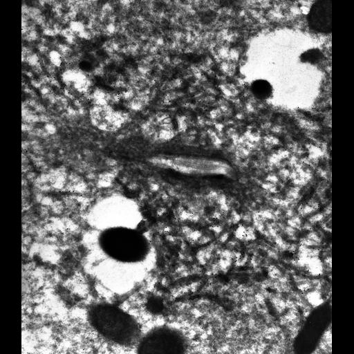
High resolution image of when the antigen was visualized with immunogold using DS-1 on thin-frozen sections the antigen could be seen to be present on the periphery of the decorated tubules. The collecting canal and smooth spongiome were not labeled with this mAb. Please see the paper for details. TEM taken on 3/22/86 by R. Allen with Zeiss 10A operating at 80kV. Neg. 12,000X. Adapted with permission from J. Cell Sci. Cells were lightly fixed with 0.25% glutaraldehyde and infiltrated with 2.3M sucrose before being frozen in liquid nitrogen and thin sectioned at a temperature of –100°C at approximately 75nm thickness. Frozen sections from these preparations were then thawed, washed, and exposed to a monoclonal primary antibody that was raised in mice or rabbit/goat and to colloidal gold-complexed goat-anti-mouse/rabbit secondary antibodies. Further details of preparation are outlined in Methods Cell Biol. 2010;96:143-73. The raw film was scanned with a Nikon Coolscan 9000ED. This image is best used for quantitative analysis. Additional information available at (http://www5.pbrc.hawaii.edu/allen/).

Biological Sources
NCBI Organism Classification
Paramecium multimicronucleatum
Cell Type
cell by organism
eukaryotic cell
Eukaryotic Protist
Ciliated Protist
Cellular Component
contractile vacuole
contractile vacuolar membrane
Biological Context
Biological Process
contractile vacuole organization
Attribution
Names
Richard Allen (University of Hawaii)
Published
J. Cell Sci. 109:229-237, 1996
Pubmed
8834807
Link
http://www5.pbrc.hawaii.edu/allen/
Citation
Digital Object Identifier (DOI)
doi:10.7295/W9CIL13121
Archival Resource Key (ARK)
ark:/b7295/w9cil13121
Grouping This image is part of a group.
Imaging
Image Type
transmission electron microscopy (TEM)
illumination by electrons
Image Mode
detection of electrons
film
Parameters Imaged
electron density
Source of Contrast
intrinsic mass distribution
stain with broad specificity
Visualization Methods
stain with broad specificity
uranyl salt
gold
Processing History
recorded image
film
unprocessed raw data
Data Qualifiers
raw, unprocessed data
suitable for spatial measurements
Sample Preparation
Methods
glutaraldehyde fixed tissue
tissue in vitreous ice embedment
microtome-sectioned tissue
monoclonal antibody
Relation To Intact Cell
microtome-sectioned tissue
Dimensions
Spatial Axis Image Size Pixel Size
X 4773px 1.25nm
Y 5438px 1.25nm