Alternate header for print version


Licensing
Attribution Non-Commercial Share Alike:This image is licensed under a Creative Commons Attribution, Non-Commercial Share Alike License. View License Deed | View Legal Code
tweet  
*CIL – Cell Image Library accession number. Please use this to reference an image.

CIL:13118*  Cite 
Description

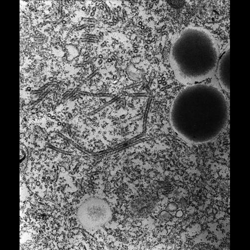
High resolution image of the decorated spongiome is composed of 50nm tubules that have a pattern of helically wound subunits attached to their cytosolic surfaces. These tubules can be up to one micrometer or more in length. Each tubule ends blindly at one end but at its opposite end it empties into a duct shared by several other tubules. This common duct empties into the smooth spongiome. TEM taken 3/13/92 by R. Allen with Zeiss 10A operating at 80kV. Neg. 19,800X. Adapted with permission from the J. Cell Sci. 108:3163-3170, 1995. The raw film was scanned with an Epson Perfection V750 Pro. This image is best used for quantitative analysis. Standard glutaraldehyde fixation followed by osmium tetroxide, dehydrated in alcohol and embedded in an epoxy resin. Microtome sections prepared at approximately 75nm thickness. Additional information available at (http://www5.pbrc.hawaii.edu/allen/).

Biological Sources
NCBI Organism Classification
Paramecium multimicronucleatum
Cell Type
cell by organism
eukaryotic cell
Eukaryotic Protist
Ciliated Protist
Cellular Component
contractile vacuolar membrane
integral to contractile vacuolar membrane
intrinsic to contractile vacuolar membrane
Biological Context
Biological Process
contractile vacuole organization
contractile vacuole discharge
Attribution
Names
Richard Allen (University of Hawaii)
Published
J. Cell Sci. 108:3163-3170, 1995
Pubmed
7593277
Link
http://www5.pbrc.hawaii.edu/allen/
Citation
Digital Object Identifier (DOI)
doi:10.7295/W9CIL13118
Archival Resource Key (ARK)
ark:/b7295/w9cil13118
Grouping This image is part of a group.
Imaging
Image Type
transmission electron microscopy (TEM)
illumination by electrons
Image Mode
detection of electrons
film
Parameters Imaged
electron density
Source of Contrast
stain with broad specificity
Visualization Methods
stain with broad specificity
osmium tetroxide
uranyl salt
lead salt
Processing History
recorded image
film
unprocessed raw data
Data Qualifiers
raw, unprocessed data
suitable for spatial measurements
Dimensions
Spatial Axis Image Size Pixel Size
X 4663px 0.76nm
Y 5540px 0.76nm