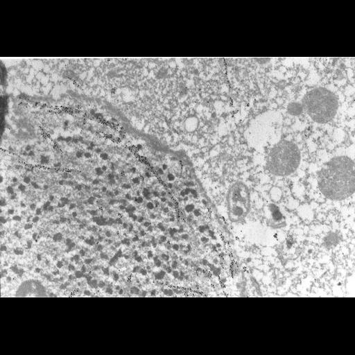
High resolution image of the macronucleus which contains microtubules that lie inside a seemingly intact nuclear envelope. Microtubules orient along the axis of macronuclear elongation in the form of small bundles. The section is immunogold labeled with anti-b-tubulin. TEM taken on 7/5/96 by R. Allen with Zeiss 10A operating at 80kV. Neg. 12,000X. were lightly fixed with 0.25% glutaraldehyde and infiltrated with 2.3M sucrose before being frozen in liquid nitrogen and thin sectioned at a temperature of –100°C at approximately 75nm thickness. Frozen sections from these preparations were then thawed, washed, and exposed to a monoclonal primary antibody that was raised in mice or rabbit/goat and to colloidal gold-complexed goat-anti-mouse/rabbit secondary antibodies. Further details of preparation are detailed in Methods Cell Biol. 2010;96:143-73. The raw film was scanned with a Nikon Coolscan 9000ED. This image is best used for quantitative analysis. Additional information available at (http://www5.pbrc.hawaii.edu/allen/).
| Spatial Axis | Image Size | Pixel Size |
|---|---|---|
| X | 5582px | 1.25nm |
| Y | 3792px | 1.25nm |