Alternate header for print version


Licensing
Public Domain: This image is in the public domain and thus free of any copyright restrictions. However, as is the norm in scientific publishing and as a matter of courtesy, any user should credit the content provider for any public or private use of this image whenever possible. Learn more
tweet  
*CIL – Cell Image Library accession number. Please use this to reference an image.

CIL:12979*  Cite 
Description

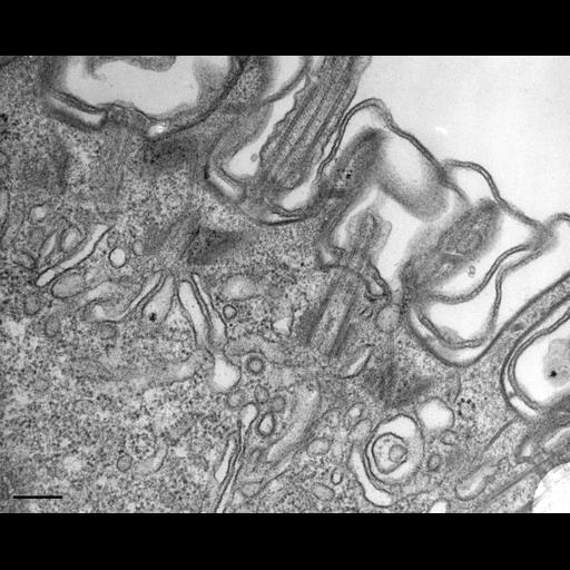
An image of a cell actively undergoing endocytosis that develops an extensive layer of vesicles and cisternae composed for the most part of smooth undecorated membrane. The uncoated cisternae are early endosomes to which uncoated vesicles that first arise as coated vesicles from the parasomal sacs fuse with the cisternae to deposit their load of receptor-mediated cargo. Smaller clathrin-coated vesicles bud from the early endosomes and these are either recycled back to the plasma membrane or move deeper into the cell as carrier vesicles after they lose their clathrin coats. TEM taken on 5/21/79 by R. Allen with Hitachi HU11A operating at 75kV. Neg. 26,250X. The negative was printed to paper and the image was scanned to Photoshop. This digitized image is available for qualitative analysis. An unprocessed, high resolution version of this image (CIL:12614) is in the library and available for quantitative analysis. Standard glutaraldehyde fixation followed by osmium tetroxide, dehydrated in alcohol and embedded in an epoxy resin. Microtome sections prepared at approximately 75nm thickness. Additional information available at (http://www5.pbrc.hawaii.edu/allen/).

Biological Sources
NCBI Organism Classification
Paramecium multimicronucleatum
Cell Type
cell by organism
eukaryotic cell
Eukaryotic Protist
Ciliated Protist
Cellular Component
cell cortex
coated pit
endosome
cell cortex
axoneme
Biological Context
Biological Process
cortical organization
endosome organization
clathrin coating of Golgi vesicle, plasma membrane to endosome targeting
Attribution
Names
Richard Allen (University of Hawaii)
Link
http://www5.pbrc.hawaii.edu/allen/
Citation
Digital Object Identifier (DOI)
doi:10.7295/W9CIL12979
Archival Resource Key (ARK)
ark:/b7295/w9cil12979
Grouping This image is part of a group.
Imaging
Image Type
transmission electron microscopy (TEM)
illumination by electrons
Image Mode
detection of electrons
film
Parameters Imaged
electron density
Source of Contrast
stain with broad specificity
Visualization Methods
stain with broad specificity
osmium tetroxide
uranyl salt
lead salt
Processing History
film
Print from negative scanned for Photoshop.
Data Qualifiers
processed data
Dimensions
Spatial Axis Image Size Pixel Size
X 2562px ——
Y 2058px ——