Alternate header for print version


Licensing
Attribution Non-Commercial Share Alike:This image is licensed under a Creative Commons Attribution, Non-Commercial Share Alike License. View License Deed | View Legal Code
tweet  
*CIL – Cell Image Library accession number. Please use this to reference an image.

CIL:12664*  Cite 
Description

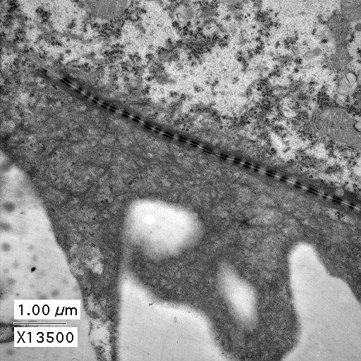
Transmission EM showing adherens junctions between cells of the enveloping layer of a 24 hr (prim-5) Zebrafish embryo. Specimens fixed in Karnofsky's (overnight at 4˚C) followed by postfixing in 4% osmium tetroxide in .2M cacodylate for 1h. The resin was LX-112 and the stain was uranyl acetate and lead citrate. This image is part of a series taken by Bryan Crawford while he was at the University of Washington. They are part of the Zebrafish--The Living Laboratory CD made available by Mark Cooper and described in Methods in Cell Biology Volume 77, 2004, Pages 439-457.

Biological Sources
NCBI Organism Classification
Danio rerio
Cellular Component
tight junction
adherens junction
Biological Context
Biological Process
embryo development
Zebrafish Anatomy
Pharyngula:Prim-5
Attribution
Names
Bryan Crawford
Link
http://www.unb.ca/fredericton/science/biology...
OTHER
Zebrafish -- The Living Laboratory
Methods in Cell Biology Volume 77, 2004, Pages 439-457
Citation
Digital Object Identifier (DOI)
doi:10.7295/W9CIL12664
Archival Resource Key (ARK)
ark:/b7295/w9cil12664
Grouping This image is part of a group.
Imaging
Image Type
recorded image
Image Mode
transmission electron microscopy (TEM)
Parameters Imaged
electron density
Source of Contrast
differences in adsorption or binding of stain
Visualization Methods
lead salt
uranyl salt
Processing History
unprocessed raw data
Data Qualifiers
raw, unprocessed data
suitable for spatial measurements
Dimensions
Spatial Axis Image Size Pixel Size
X 512px 0.0128µm
Y 512px 0.0128µm