Alternate header for print version


Licensing
This image is copyright protected. Any public or private use of this image is subject to prevailing copyright laws. Please contact the content provider of this image for permission requests.
tweet  
*CIL – Cell Image Library accession number. Please use this to reference an image.

CIL:12655*  Cite 
Description

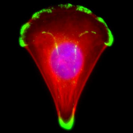
Human primary keratinocyte spreading on an asymmetric "arc"-shaped collagen I micropattern. The cells are stained for the focal adhesion protein vinculin (green, Alexa488), F-Actin (red, TRITC phalloidin) and the nucleus (blue, DAPI). The polymer brush micropatterning technique used is described in Gautrot et al. Biomaterials 2010, 31, 5030-5040. Images were acquired on a Leica DMI4000 using a 63X, NA: 1.25 and a Leica DFC 340 FX camera with 17 ms exposure for blue; 117 ms for green, and 159 ms for red.

Biological Sources
NCBI Organism Classification
Homo sapiens
Cell Type
keratinocyte
Cellular Component
focal adhesion
actin cytoskeleton
nucleus
Biological Context
Biological Process
substrate adhesion-dependent cell spreading
Attribution
Names
Julien Gautrot
Citation
Digital Object Identifier (DOI)
doi:10.7295/W9CIL12655
Archival Resource Key (ARK)
ark:/b7295/w9cil12655
Imaging
Image Type
recorded image
Image Mode
fluorescence microscopy
Parameters Imaged
fluorescence emission
Source of Contrast
distribution of a specific protein
Visualization Methods
4',6-diamidino-2-phenylindole (DAPI)
Alexa Fluor 488
Tetramethyl rhodamine (TRITC)
phalloidin
primary antibody plus labeled secondary antibody
Processing History
color combine
Data Qualifiers
raw, unprocessed data
suitable for spatial measurements
Sample Preparation
Methods
formaldehyde fixed tissue
permeabilized tissue
Relation To Intact Cell
dispersed cells in vitro
Dimensions
Spatial Axis Image Size Pixel Size
X 380px 0.1µm
Y 520px 0.1µm
Channel Wavelength
1 DAPI, Alexa488, TRITCnm