Alternate header for print version


Licensing
Public Domain: This image is in the public domain and thus free of any copyright restrictions. However, as is the norm in scientific publishing and as a matter of courtesy, any user should credit the content provider for any public or private use of this image whenever possible. Learn more
tweet  
*CIL – Cell Image Library accession number. Please use this to reference an image.

CIL:12627*  Cite 
Description

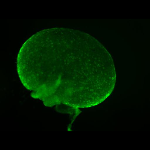
Fluorescent image of a mouse ovary labeled with an antibody to vasa (involved in oocyte differentiation and polarity) which is localized in the oocytes within the ovary. The sample was fixed in paraformaldehyde for subsequent immunolabeling with an antibody to vasa and a secondly antibody conjugated to Alexa 488. The image was captured with a 4MP RGB/gray SPOT digital camera mounted on a Leica MZII Pursuit fluorescent stereoscopic microscope.

Biological Sources
NCBI Organism Classification
Mus musculus
Cell Line
Ovary
Cellular Component
vasa
Biological Context
Biological Process
oocyte maturation
Attribution
Names
Prue Talbot
Citation
Digital Object Identifier (DOI)
doi:10.7295/W9CIL12627
Archival Resource Key (ARK)
ark:/b7295/w9cil12627
Sample Preparation
Methods
formaldehyde fixed tissue
Relation To Intact Cell
whole mounted tissue
Dimensions
Spatial Axis Image Size Pixel Size
X 800px ——
Y 600px ——