Alternate header for print version


Licensing
Public Domain: This image is in the public domain and thus free of any copyright restrictions. However, as is the norm in scientific publishing and as a matter of courtesy, any user should credit the content provider for any public or private use of this image whenever possible. Learn more
tweet  
*CIL – Cell Image Library accession number. Please use this to reference an image.

CIL:12625*  Cite 
Description

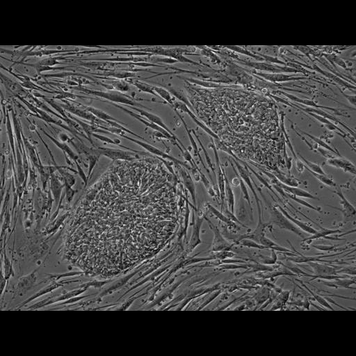
Human embryonic stem cell colonies. This phase contrast image shows two human embryonic stem cell colonies growing on a feeder layer of mouse embryonic fibroblasts. The stem cells actually grow in between the fibroblasts. The feeder layer helps provide nutrition for the stem cells as well as factors that keep them from differentiating. This is a live cell preparation that was imaged using a Nikon inverted Eclipse microscope.

Biological Sources
NCBI Organism Classification
Homo sapiens
Mus musculus
Cell Type
stem cell
fibroblast
Cellular Component
cell
Biological Context
Biological Process
stem cell proliferation
Attribution
Names
Sabrina Lin
Prue Talbot
OTHER
Stem Cell Center University of California, Riverside
Citation
Digital Object Identifier (DOI)
doi:10.7295/W9CIL12625
Archival Resource Key (ARK)
ark:/b7295/w9cil12625
Grouping This image is part of a group.
Imaging
Image Type
recorded image
Image Mode
phase contrast microscopy
Parameters Imaged
elastic scattering of photons
Source of Contrast
differences in amount of elastic light scattering
Visualization Methods
visualization of contiguous regions
Processing History
unprocessed raw data
Sample Preparation
Methods
living tissue
Relation To Intact Cell
dispersed cells in vitro
Dimensions
Spatial Axis Image Size Pixel Size
X 1392px ——
Y 1040px ——