Alternate header for print version


Licensing
Public Domain: This image is in the public domain and thus free of any copyright restrictions. However, as is the norm in scientific publishing and as a matter of courtesy, any user should credit the content provider for any public or private use of this image whenever possible. Learn more
tweet  
*CIL – Cell Image Library accession number. Please use this to reference an image.

CIL:12618*  Cite 
Description

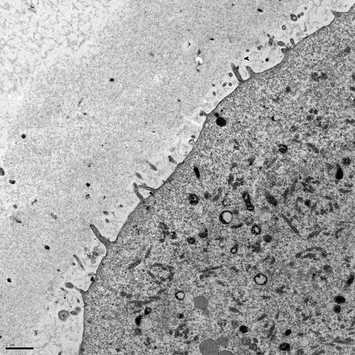
Surface of a mouse oocyte This transmission electron micrograph shows the cortex of a mouse oocyte that was undergoing fertilization. The sample was fixed using glutaraldehyde and osmium tetroxide, embedded in plastic, sectioned, and stained with uranyl acetate and lead citrate. Microvilli are present on the oocyte's surface and the zona pellucida is visible surrounding the oocyte. The image was taken with a Phillips 500 transmission electron microscope. The scale bar is 1

Biological Sources
NCBI Organism Classification
Mus musculus
Cell Type
oocyte
Cellular Component
cell cortex
microvillus
zona pellucida
Biological Context
Biological Process
fertilization
Attribution
Names
Prue Talbot
Citation
Digital Object Identifier (DOI)
doi:10.7295/W9CIL12618
Archival Resource Key (ARK)
ark:/b7295/w9cil12618
Imaging
Image Type
recorded image
Image Mode
transmission electron microscopy (TEM)
Parameters Imaged
electron density
Source of Contrast
differences in adsorption or binding of stain
Visualization Methods
uranyl salt
lead salt
Processing History
unprocessed raw data
Data Qualifiers
raw, unprocessed data
suitable for spatial measurements
Dimensions
Spatial Axis Image Size Pixel Size
X 2048px 0.0067µm
Y 2048px 0.0067µm