Alternate header for print version


Licensing
Public Domain: This image is in the public domain and thus free of any copyright restrictions. However, as is the norm in scientific publishing and as a matter of courtesy, any user should credit the content provider for any public or private use of this image whenever possible. Learn more
tweet  
*CIL – Cell Image Library accession number. Please use this to reference an image.

CIL:12604*  Cite 
Description

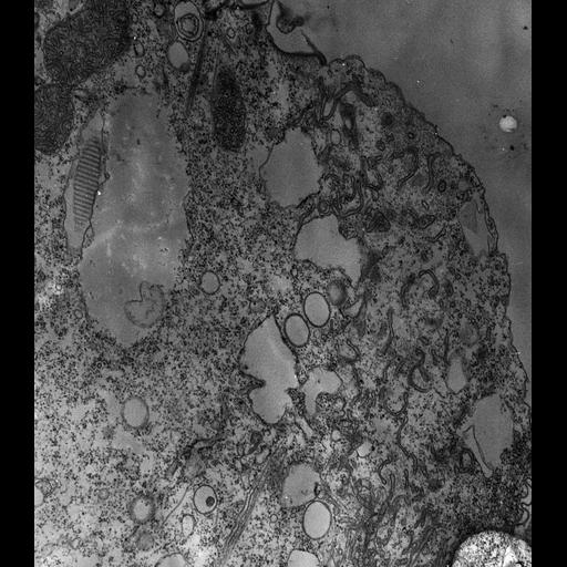
High resolution image of a colchicine-treated cell, used in an attempt to disrupt its microtubules. We found that microtubules already formed are not disrupted in Paramecium multimicronucleatum. This open cytoproct shows the microfilaments around the retrieved membrane vesicles. The larger vesicles appear to be expanded ER as ribosomes can be seen on their cytosolic face. A long membrane tubule appears to be bound to a microtubular ribbon that may have its origin at the cytopharynx. TEM taken on 1/16/73 by R. Allen with Hitachi HU11A operating at 60kV. Neg. 8,100X.

Technical Details

The raw film was scanned with an Epson Perfection V750 Pro. This image is best used for quantitative analysis. Standard glutaraldehyde fixation followed by osmium tetroxide, dehydrated in alcohol and embedded in an epoxy resin. Microtome sections prepared at approximately 75nm thickness. Additional information available at (http://www5.pbrc.hawaii.edu/allen/).

Biological Sources
NCBI Organism Classification
Paramecium multimicronucleatum
Cell Type
cell by organism
eukaryotic cell
Eukaryotic Protist
Ciliated Protist
Cellular Component
cytoproct
cell cortex
microtubule
Biological Context
Biological Process
microtubule cytoskeleton organization
Attribution
Names
Richard Allen (University of Hawaii)
Link
http://www5.pbrc.hawaii.edu/allen/
Citation
Digital Object Identifier (DOI)
doi:10.7295/W9CIL12604
Archival Resource Key (ARK)
ark:/b7295/w9cil12604
Grouping This image is part of a group.
Imaging
Image Type
transmission electron microscopy (TEM)
illumination by electrons
Image Mode
detection of electrons
film
Parameters Imaged
electron density
Source of Contrast
stain with broad specificity
Visualization Methods
stain with broad specificity
osmium tetroxide
uranyl salt
lead salt
Processing History
recorded image
film
unprocessed raw data
Data Qualifiers
raw, unprocessed data
suitable for spatial measurements
Dimensions
Spatial Axis Image Size Pixel Size
X 4958px 1.6nm
Y 5646px 1.6nm