Licensing

Pd
Public Domain: This image is in the public domain and thus free of any copyright restrictions. However, as is the norm in scientific publishing and as a matter of courtesy, any user should credit the content provider for any public or private use of this image whenever possible. Learn more
Comments: Powered by Pnyxe

Description

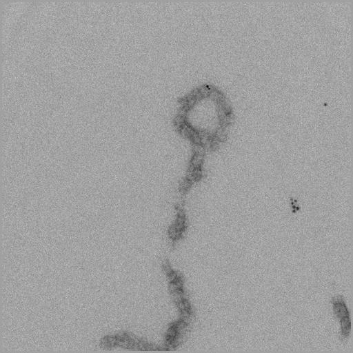
Telomeres isolated from chicken erythrocyte nuclei following restriction endonuclease treatment, and imaged by transmission EM after uranyl acetate negative staining. Specimen was also labeled with immunogold using an anti-TRF1 primary antibody. Telomere chromatin adopts a characteristic looped conformation which likely aids in protection from the rapid degradation that occurs with free DNA ends.

Technical Details

Image recorded with FEI Tecnai 12 TEM at 100KV and recorded on 2048x2048 CCD with 2x binning. See also: T Nikitina and C L Woodcock 2004 Closed chromatin loops at the ends of chromosomes. J Cell Biol 166:161-165.

Biological Sources

NCBI Organism Classification
Gallus gallus gallus
Cell Type
erythrocyte
Cellular Component
chromosome, telomeric region
chromatin
nucleosome

Biological Context

Biological Process
chromatin assembly
chromatin organization
Molecular Function
structural constituent of chromatin

Attribution

Names
Christopher Woodcock
Tatiana Nikitina

Imaging

Image Type
charge coupled device (CCD)
Imaging Mode
transmission electron microscopy (TEM)
Parameters Imaged
electron density
Source of Contrast
differences in adsorption or binding of stain
Visualization Methods
negative staining
gold
uranyl salt
Processing History
unprocessed raw image

Sample Preparation

Methods
glutaraldehyde fixed tissue
Relation To Intact Cell
isolated subcellular component

Dimensions

Spatial Axis Image Size Pixel Size
X 1024px 0.65nm
Y 1024px 0.65nm
*CIL – Cell Image Library accession number. Please use this to reference an image.