Alternate header for print version


Licensing
Attribution Only: This image is licensed under a Creative Commons Attribution License. View License Deed | View Legal Code
tweet  
*CIL – Cell Image Library accession number. Please use this to reference an image.

CIL:12597*  Cite 
Description

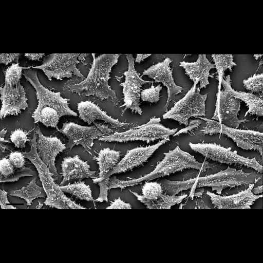
Scanning electron micrograph montage of cultured HeLa cells. This image is a montage of six overlapping fields of view, each photographed at 2,231x with a 17.8mm working distance and an accelerating voltage of 12.5kV. The cells were grown to confluence on Thermanox® coverslips and fixed with glutaraldehyde and osmium tetroxide, then critical point dried. These cells were imaged using an FEI Quanta 200 scanning electron microscope. Rounded, non-adherent cells are most likely mitotic, and the two adjacent, spherical cells near the lower left corner have almost completed cytokinesis. Scale bar = 10µm.

Biological Sources
NCBI Organism Classification
Homo sapiens
Cell Type
epithelial cell
Cell Line
HeLa
Cellular Component
cell
cell surface
Biological Context
Biological Process
cytokinesis after mitosis
Attribution
Names
Anna Baccei
Marian Rice
OTHER
Mount Holyoke College
Citation
Digital Object Identifier (DOI)
doi:10.7295/W9CIL12597
Archival Resource Key (ARK)
ark:/b7295/w9cil12597
Imaging
Image Type
recorded image
montage
Image Mode
scanning electron microscopy (SEM)
Parameters Imaged
elastic scattering of electrons
Source of Contrast
differences in adsorption or binding of stain
Visualization Methods
osmium tetroxide
Processing History
unprocessed raw data
montage
Data Qualifiers
raw, unprocessed data
suitable for spatial measurements
Sample Preparation
Methods
glutaraldehyde fixed tissue
critical_point dried specimen
Relation To Intact Cell
dispersed cells in vitro
Dimensions
Spatial Axis Image Size Pixel Size
X 2674px 0.056µm
Y 1587px 0.056µm