Alternate header for print version


Licensing
Public Domain: This image is in the public domain and thus free of any copyright restrictions. However, as is the norm in scientific publishing and as a matter of courtesy, any user should credit the content provider for any public or private use of this image whenever possible. Learn more
tweet  
*CIL – Cell Image Library accession number. Please use this to reference an image.

CIL:12595*  Cite 
Description

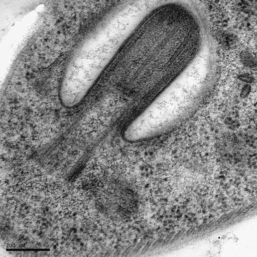
The flagellar pocket of a Trypanosoma brucei bloodstream form cell, with short flagellum. The image shows a new flagellum starting to grow from the flagellar pocket, in preparation for the next cell division. Cells were high pressure frozen and freeze substituted (1hr freeze substitution in 2% uranyl acetate in acetone and 8% methanol). An 80nm thick section was stained for 5 minutes with uranyl acetate, one minute with lead citrate and viewed using a Philips CM100 transmission electron microscope. The image was captured with an Advantage HS-B camera (2000 x 2000 pixels; AMT, Danvers, MA, USA). Contrast adjusted using Photoshop. Magnification indicated by scale bar.

Biological Sources
NCBI Organism Classification
Trypanosoma brucei
Cell Type
cell by organism
eukaryotic cell
Eukaryotic Protist
Flagellated Protist
Cellular Component
microtubule basal body
axoneme
axoneme part
cortical microtubule, transverse to long axis
Attribution
Names
Johanna Hoog
Citation
Digital Object Identifier (DOI)
doi:10.7295/W9CIL12595
Archival Resource Key (ARK)
ark:/b7295/w9cil12595
Imaging
Image Type
transmission electron microscopy (TEM)
illumination by electrons
Image Mode
detection of electrons
electron bombardment CCD (EBCCD)
Parameters Imaged
electron density
Source of Contrast
stain with broad specificity
Visualization Methods
stain with broad specificity
uranyl salt
lead salt
Processing History
recorded image
electron bombardment CCD (EBCCD)
Contrast enhanced in Photoshop.
Data Qualifiers
processed data
suitable for spatial measurements
Sample Preparation
Methods
freeze substitution
microtome-sectioned tissue
Relation To Intact Cell
microtome-sectioned tissue
Dimensions
Spatial Axis Image Size Pixel Size
X 2048px ——
Y 2048px ——