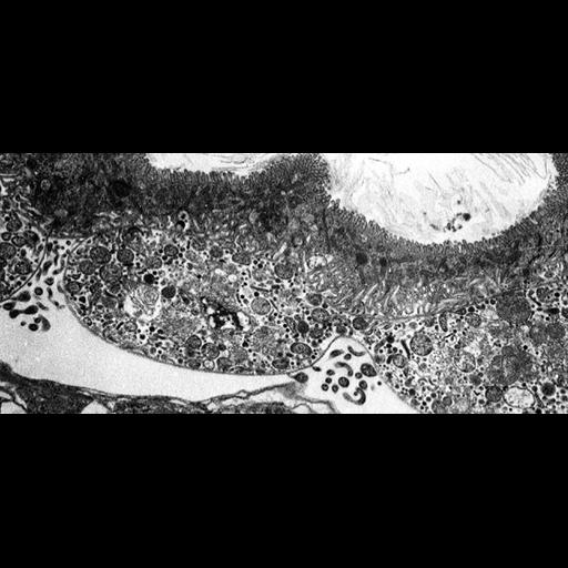
Electron micrograph of corn stunt spiroplasma (CSS) between 2 split layers of the basal lamina surrounding the filter chamber (part of the alimentary canal) and in the hemolymph of a vector leafhopper D. gelbus. Note the mainly pleiomorphic forms in host cells and the filamentous/spiral forms in the hemolymph. CIL:19131, from the same image group, is a labeled version of this image. CSS is a wall-less procaryote that causes corn stunt disease and is biologically transmitted by some leafhopper species, e.g. Dalbulus maidis and D. gelbus (Hemiptera, Cicadellidae). Spiroplasma cells are limited by a unit membrane with no cell wall. They can be quasi-spherical, pleiomorphic or maintain a filamentous/spiral form. CSS multiplies in both plant and insect hosts and is transmitted with salivary secretions during feeding of the vector on new host plants. Additional images from this contributor are available in the Library.
Detailed methods: Tissue was processed for TEM by fixation in glutaraldehyde and osmium tetroxide, embedded in Spurr's medium. Thin sections were stained with uranyl acetate and lead citrate, and examined by a Philips-201 TEM (non-digital camera).
| Spatial Axis | Image Size | Pixel Size |
|---|---|---|
| X | 877px | 0.0149µm |
| Y | 404px | 0.0149µm |