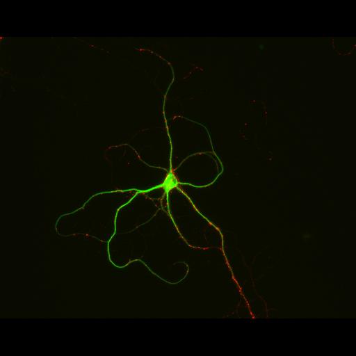
Presynaptic contacts are abundant on both cell bodies and the dendritic arbor of cultured hippocampal neurons after 15 days in vitro. Neurons were immunostained for MAP2, a microtubule associated protein localized to dendrites (green) and synapsin I (red), a presynaptic vesicle protein. With the exception of the immunostained presynaptic terminals, axons are not visible in this preparation. A relatively low power objective (20X) was used to capture the entire dendritic arbor of individual cells. Detailed methods: Embryonic rat hippocampal neurons were prepared as previously described (see Kaech and Banker, 2006, Nat Protoc). Cells were prepared for fluorescent staining as previously described (Withers and Banker, 1998, in Culturing Nerve Cells, MIT Press). Briefly, cells were fixed (4% formaldehyde, 4% sucrose in phosphate buffered saline, pH 7.4), permeabilized with 0.25% Triton and immunostained for MAP2 (monoclonal HM2, Sigma, with Alexa 488 conjugated secondary, excitation, 494, emission, 519 [Invitrogen, Molecular Probes]) and synapsin I (from P. DeCamilli, with DyLight549 conjugated secondary, excitation, 555, emission, 568, [Jackson Immunoresearch]). Images were acquired with a Leica DMRA microscope with a 20X (HC Fluotar, NA 0.5) lens, Photometrics CoolSnap ES CCD camera and MetaMorph software. Individual images of each fluorophore were colorized and assembled as a a stack file using MetaMorph software.
| Spatial Axis | Image Size | Pixel Size |
|---|---|---|
| X | 1300px | 0.339µm |
| Y | 1030px | 0.339µm |