Alternate header for print version


Licensing
Public Domain: This image is in the public domain and thus free of any copyright restrictions. However, as is the norm in scientific publishing and as a matter of courtesy, any user should credit the content provider for any public or private use of this image whenever possible. Learn more
tweet  
*CIL – Cell Image Library accession number. Please use this to reference an image.

CIL:12340*  Cite 
Description

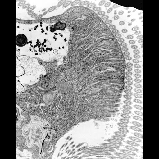
A view of the cytopharynx and its nascent food vacuole forming region. The flattened pharyngeal vesicles that may contain monolayers or bilayers of lipids seem to be equivalent to the discoidal vesicles of Paramecium that provide membrane for food vacuole growth. They approach and fuse with the cytopharyngeal membrane between the lamellae (cytopharyngeal ribbons). TEM taken on 7/29/67 by R. Allen with Philips 200 operating at 60kV. Neg. 9,000X. Bar = 0.5µm.

Technical Details

Standard glutaraldehyde fixation followed by osmium tetroxide, dehydrated in alcohol, and embedded in an epoxy resin. Microtome sections prepared at approximately 75nm thickness. A print of the negative was scanned and processed in Photoshop. This image is best used for qualitative analysis. There is a high resolution version of this image in the library (CIL:39114) which is available for quantitative analysis. Additional information available at (http://www5.pbrc.hawaii.edu/allen/).

Biological Sources
NCBI Organism Classification
Euplotes sp.
Cell Type
cell by organism
eukaryotic cell
Eukaryotic Protist
Ciliated Protist
Cell Line
Euplotes sp. from Carolina Biological Supply
Cellular Component
oral apparatus
cortical cytoskeleton
Attribution
Names
Richard Allen
Link
http://www5.pbrc.hawaii.edu/allen/
Citation
Digital Object Identifier (DOI)
doi:10.7295/W9CIL12340
Archival Resource Key (ARK)
ark:/b7295/w9cil12340
Grouping This image is part of a group.
Imaging
Image Type
transmission electron microscopy (TEM)
illumination by electrons
Image Mode
detection of electrons
film
Parameters Imaged
electron density
Source of Contrast
stain with broad specificity
Visualization Methods
stain with broad specificity
osmium tetroxide
uranyl salt
lead salt
Processing History
recorded image
film
Print from negative scanned for Photoshop.
Data Qualifiers
processed data
suitable for spatial measurements
Dimensions
Spatial Axis Image Size Pixel Size
X 2406px ——
Y 3000px ——