Licensing

Attribution_nc_sa
Attribution Non-Commercial Share Alike:This image is licensed under a Creative Commons Attribution, Non-Commercial Share Alike License. View License Deed | View Legal Code
Comments: Powered by Pnyxe

Description

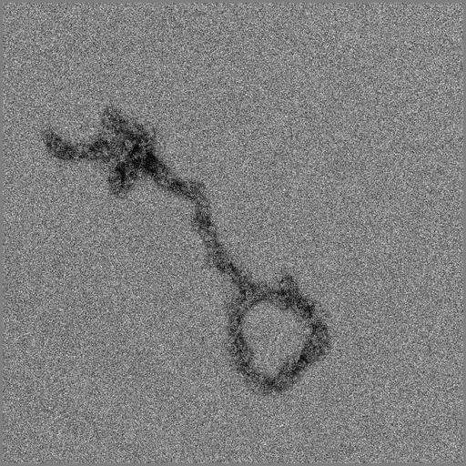
Telomeres were isolated from chicken erythrocyte nuclei following restriction endonuclease treatment, and imaged by transmission EM after uranyl acetate negative staining. Like its DNA counterpart, telomeric chromatin has have a looped conformation, which likely aids in protection from the rapid degradation that occurs with free DNA ends.

Technical Details

The image was recorded with a FEI Tecnai 12 TEM at 100KV and recorded on a TVIPS 2048x2048 CCD with 2x binning. See Nikitina T, Woodcock CL. 2004. Closed chromatin loops at the ends of chromosomes. J Cell Biol.166:161-5.

Biological Sources

NCBI Organism Classification
Gallus gallus gallus
Cell Type
erythrocyte
Cellular Component
chromosome, telomeric region

Biological Context

Biological Process
chromatin assembly
chromosome organization
Molecular Function
structural constituent of chromatin

Attribution

Name
Chris Woodcock
Published
Nikitina T, Woodcock CL. 2004. Closed chromatin loops at the ends of chromosomes. J Cell Biol.166:161-5.
Pubmed
2172301

Imaging

Image Type
charge coupled device (CCD)
Imaging Mode
transmission electron microscopy (TEM)
Parameters Imaged
electron density
Source of Contrast
differences in adsorption or binding of stain
Visualization Methods
uranyl salt
Processing History
unprocessed raw image

Sample Preparation

Methods
glutaraldehyde fixed tissue
Relation To Intact Cell
isolated subcellular component

Dimensions

Spatial Axis Image Size Pixel Size
X 2048px 0.454nm
Y 2048px 0.454nm
*CIL – Cell Image Library accession number. Please use this to reference an image.