Alternate header for print version


Licensing
Public Domain: This image is in the public domain and thus free of any copyright restrictions. However, as is the norm in scientific publishing and as a matter of courtesy, any user should credit the content provider for any public or private use of this image whenever possible. Learn more
tweet  
*CIL – Cell Image Library accession number. Please use this to reference an image.

CIL:12075*  Cite 
Description

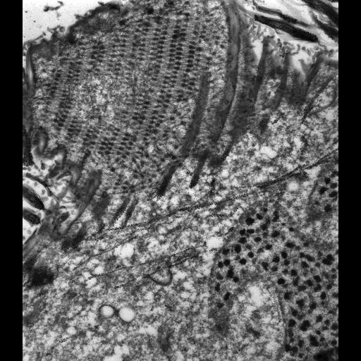
High resolution view of the bundles of microtubules that arise from along the elaborately woven blanket of the filamentous reticulum that covers the ribbed wall of the right side of the buccal cavity. A bundle forms beneath the bottom of each rib of the ribbed wall and runs parallel to the depression of the rib. These bundles then extend beyond the posterior terminus of the buccal cavity where they are then collectively called the postoral fiber. In addition single microtubules seem to lie deeper in the cytosol at approximately right angles to the bundles. TEM taken on 7/5/96 by R. Allen with Zeiss 10A operating at 80kV. Neg. 9,780X.Cells were lightly fixed with 0.25% glutaraldehyde and infiltrated with 2.3M sucrose before being frozen in liquid nitrogen and thin sectioned at a temperature of –100°C at approximately 75nm thickness. Frozen sections from these preparations were then thawed, washed, and exposed to a monoclonal primary antibody that was raised in mice or rabbit/goat and to colloidal gold-complexed goat-anti-mouse/rabbit secondary antibodies. Further details of preparation are detailed in Methods Cell Biol. 2010;96:143-73. The raw film was scanned with a Nikon Coolscan 9000ED. This image is best used for quantitative analysis. Additional information available at (http://www5.pbrc.hawaii.edu/allen/).

Biological Sources
NCBI Organism Classification
Paramecium tetraurelia
Cell Type
cell by organism
eukaryotic cell
Eukaryotic Protist
Ciliated Protist
Cellular Component
cell cortex
cortical microtubule cytoskeleton
cortical microtubule
oral apparatus
macronucleus
Attribution
Names
Richard Allen (University of Hawaii)
Link
http://www5.pbrc.hawaii.edu/allen/
Citation
Digital Object Identifier (DOI)
doi:10.7295/W9CIL12075
Archival Resource Key (ARK)
ark:/b7295/w9cil12075
Grouping This image is part of a group.
Imaging
Image Type
transmission electron microscopy (TEM)
illumination by electrons
Image Mode
detection of electrons
film
Parameters Imaged
electron density
Source of Contrast
intrinsic mass distribution
stain with broad specificity
Visualization Methods
stain with broad specificity
uranyl salt
gold
Processing History
recorded image
film
unprocessed raw data
Data Qualifiers
raw, unprocessed data
suitable for spatial measurements
Sample Preparation
Methods
glutaraldehyde fixed tissue
tissue in vitreous ice embedment
microtome-sectioned tissue
monoclonal antibody
Relation To Intact Cell
microtome-sectioned tissue
Dimensions
Spatial Axis Image Size Pixel Size
X 5314px 1.4nm
Y 6062px 1.4nm