Alternate header for print version


Licensing
Attribution Non-Commercial; No Derivatives:This image is licensed under a Creative Commons Attribution, Non-Commercial, No Derivatives License. View License Deed | View Legal Code
tweet  
*CIL – Cell Image Library accession number. Please use this to reference an image.

CIL:11832*  Cite 
Description

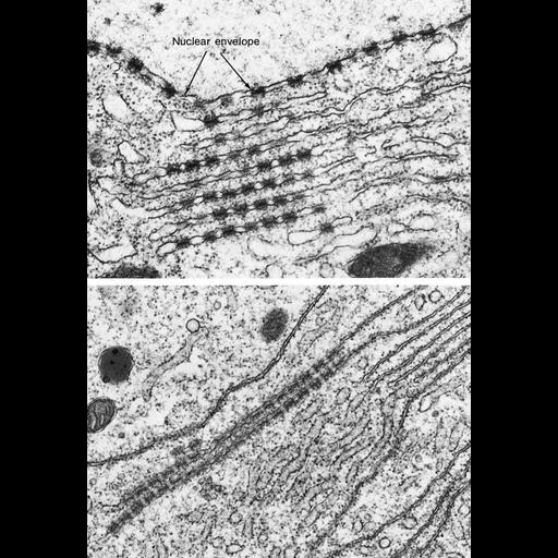
Transmission electron micrograph of stacked annulate lamellae from the oocyte of the sea urchin Arbacia. Annulate lamellae consist of sheets of membrane containing closely packed structures resembling nuclear pores. Micrographs courtesy of Susumu Ito, Figures 161 (upper) and 162 (lower) from Chapter 4 (Nucleus) of 'The Cell, 2nd Ed.' by Don W. Fawcett M.D. A PDF copy of the accompanying chapter is available on the ASCB's BioEDUCATE website.

Biological Sources
NCBI Organism Classification
Arbacia
Cell Type
oocyte
Cellular Component
annulate lamellae
nuclear envelope
nuclear pore
endoplasmic reticulum
Biological Context
Biological Process
nucleus organization
nuclear pore organization
Attribution
Names
Susumu Ito
Published
ISBN 0721635849
Published
from: Don W. Fawcett M.D ‘The Cell’ 2nd edition, W.B. Saunders Chapter 4 (Nucleus)
Link
ASCB BioEDUCATE program
ASCB Bioeducate Program
Citation
Digital Object Identifier (DOI)
doi:10.7295/W9CIL11832
Archival Resource Key (ARK)
ark:/b7295/w9cil11832
Grouping This image is part of a group.
Imaging
Image Type
film
Image Mode
transmission electron microscopy (TEM)
detection of electrons
Parameters Imaged
electron density
Source of Contrast
differences in adsorption or binding of stain
Visualization Methods
uranyl salt
osmium tetroxide
lead salt
Processing History
Image extracted from .pdf file of 'The Cell'
Data Qualifiers
processed data
Sample Preparation
Methods
tissue in plastic embedment
osmium tetroxide fixed tissue
Relation To Intact Cell
microtome-sectioned tissue
Dimensions
Spatial Axis Image Size Pixel Size
X 875px ——
Y 1271px ——