Licensing

Attribution_by_nc_nd
Attribution Non-Commercial; No Derivatives:This image is licensed under a Creative Commons Attribution, Non-Commercial, No Derivatives License. View License Deed | View Legal Code
Comments: Powered by Pnyxe

Description

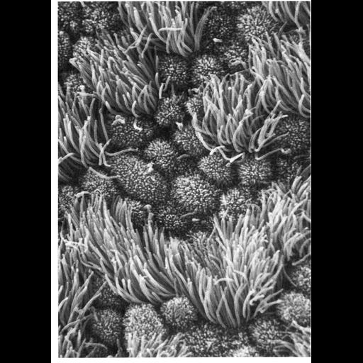
Figure 316 from Chapter 13 (Cilia and Flagella) of 'The Cell' by Don W. Fawcett M.D. The differences in size and shape of cilia and microvilli are well illustrated by scanning micrographs of the lumenal surface of the epithelium lining the mammalian oviduct. The tufts of cilia associated with individual ciliated cells project several microns above the convex apices of nonciliated cells covered with short microvilli. The number of ciliated cells in this epithelium is under hormonal control by estrogens. A copy of the chapter is available on the ASCB's BioEDUCATE website.

Biological Sources

NCBI Organism Classification
Homo sapiens
(human)
Cell Type
ciliated epithelial cell
oviduct epithielial cell
Cellular Component
cilium
microvillus

Biological Context

Biological Process
cilia organization
microvillus organization
Human Development Anatomy
homo sapiens

Attribution

Names
Don W. Fawcett
Gaddum-Rosse
Blandau
Tiersch
Publisheds
Am. J. Anat. 138:269-275, 1973
ISBN 0721635849
Link
http://www.ascb.org/bioeducate/Fawcett...

Grouping

This image is part of a group.
Data Qualifiers
processed data

Dimensions

Spatial Axis Image Size Pixel Size
X 910px ——
Y 1268px ——
*CIL – Cell Image Library accession number. Please use this to reference an image.