Alternate header for print version


Licensing
This image is copyright protected. Any public or private use of this image is subject to prevailing copyright laws. Please contact the content provider of this image for permission requests.
tweet  
*CIL – Cell Image Library accession number. Please use this to reference an image.

CIL:11564*  Cite 
Description

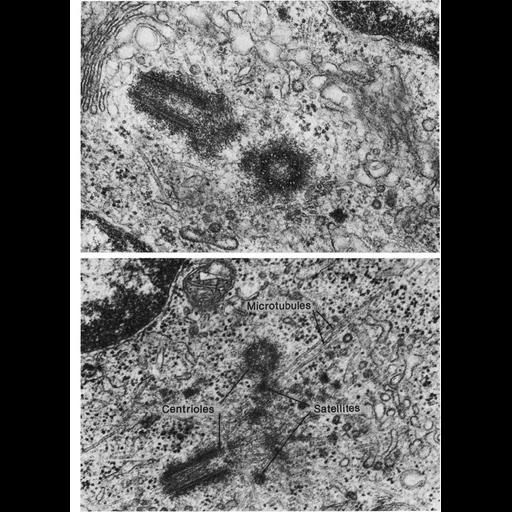
Figs. 309 & 310 from Don Fawcett's Chapter 12 (Centrioles). The plane of a thin section only rarely happens to coincide with the long axis of both members of a pair of centrioles. It is more common for one to be cut longitudinally and the other transversely, as in the accompanying micrographs. In both of these examples, the centrioles are oriented at right angles even though they are some distance apart. The pair of centrioles in the upper micrograph on the facing page is closely associated with the Golgi apparatus and occupies a concavity in the nucleus. This is a common relationship in the hemopoietic cell line and in other cell types as well. The lower figure illustrates the special character of the pericentriolar cytoplasm which often contains numerous satellites. The labeled microtubules end in one of the satellites. A copy of the chapter is available on the ASCB's BioEDUCATE website.

Biological Sources
NCBI Organism Classification
Cavia porcellus
ascites tumor cell
Cell Type
ascites tumor cell
Cellular Component
centriole
pericentriolar material
microtubule
centriole satellites
Biological Context
Biological Process
centriole satellites
Attribution
Names
Don W. Fawcett
Guy de The
Published
ISBN 0721635849
Published
J. Cell Biol. 23:265-275, 1964
Pubmed
14222814
Link
http://www.ascb.org/bioeducate/FawcettTheCell...
Citation
Digital Object Identifier (DOI)
doi:10.7295/W9CIL11564
Archival Resource Key (ARK)
ark:/b7295/w9cil11564
Grouping This image is part of a group.
Imaging
Image Type
transmission electron microscopy (TEM)
illumination by electrons
Image Mode
detection of electrons
film
Parameters Imaged
electron density
Source of Contrast
stain with broad specificity
Visualization Methods
stain with broad specificity
osmium tetroxide
uranyl salt
lead salt
Processing History
recorded image
film
Print from negative scanned for Photoshop.
Data Qualifiers
processed data
Dimensions
Spatial Axis Image Size Pixel Size
X 914px ——
Y 1260px ——