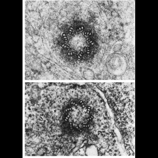
Figs. 303 & 304 from Don Fawcett's Chapter 12 (Centrioles). In transverse section the cylindrical wall of the centriole is made up of nine longitudinally oriented triplet microtubules. A dense material of unknown nature occupies the interstices between the triplets. Each triplet is at a constant angle of about 40 degrees to its respective tangent. A pattern is thus formed that resembles the vanes of a turbine or the charges of a pyrotechnic "pinwheel." The innermost microtubule of each triplet has a circular cross section and is designated subunit a and the other two b and c. The latter two each share a segment of the wall of the adjacent microtubule and therefore have a C-shaped profile instead of a circular cross section. Subunit a has two short diverging projections resembling the arms on the doublets of the ciliary axoneme. One of these is directed inward along a radius and appears to have a free end pointing toward the center of the centriole. The other projection connects with subunit c of the next triplet. The successive triplets are thus linked together a to c around the circumference of the centriole by a series of linear densities. It is not known whether these latter correspond to the dynein arms or nexin links of flagellar axonemes. A copy of the chapter is available on the ASCB's BioEDUCATE website.
| Spatial Axis | Image Size | Pixel Size |
|---|---|---|
| X | 918px | —— |
| Y | 1288px | —— |