Alternate header for print version


Licensing
Attribution Non-Commercial; No Derivatives:This image is licensed under a Creative Commons Attribution, Non-Commercial, No Derivatives License. View License Deed | View Legal Code
tweet  
*CIL – Cell Image Library accession number. Please use this to reference an image.

CIL:11439*  Cite 
Description

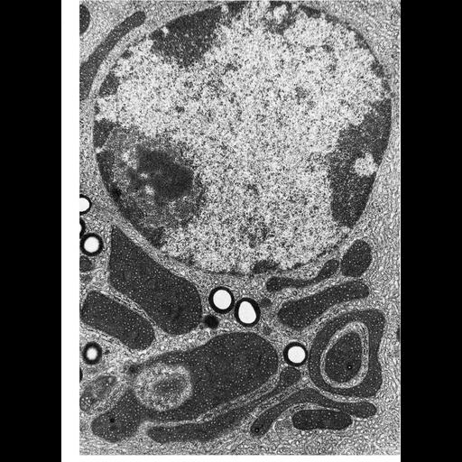
Mitochondria can show tremendous variation in size, even within a single cell. Here, a huge spherical mitochondria of about 6-7 µm in diameter is adjacent to small elongate mitochondria 0.5 µm wide in a testicular Leydig cell of the ground squirrel Citellus lateralis. This image is courtesy of Jeffrey Pudney, and appears as Figure 247 in Chapter 7 (Mitochondria) of 'The Cell, 2nd Ed.' by Don W. Fawcett M.D. A PDF copy of the accompanying chapter is available on the ASCB’s BioEDUCATE website.

Biological Sources
NCBI Organism Classification
Spermophilus citellus
Cell Type
Leydig cell
Cellular Component
mitochondrion
mitochondrial crista
mitochondrial outer membrane
mitochondrial inner membrane
Biological Context
Biological Process
cellular respiration
Attribution
Names
Jeffrey Pudney
Published
ISBN 0721635849
Link
Fawcett's "The Cell" 2nd Ed.
Citation
Digital Object Identifier (DOI)
doi:10.7295/W9CIL11439
Archival Resource Key (ARK)
ark:/b7295/w9cil11439
Grouping This image is part of a group.
Imaging
Image Type
transmission electron microscopy (TEM)
Image Mode
illumination by electrons
detection of electrons
Parameters Imaged
electron density
Source of Contrast
stain with broad specificity
Data Qualifiers
processed data
Sample Preparation
Methods
crosslinking-fixative fixed tissue
embedded tissue
Relation To Intact Cell
microtome-sectioned tissue
Dimensions
Spatial Axis Image Size Pixel Size
X 934px ——
Y 1272px ——