Licensing

Attribution_nc_sa
Attribution Non-Commercial Share Alike:This image is licensed under a Creative Commons Attribution, Non-Commercial Share Alike License. View License Deed | View Legal Code
Comments: Powered by Pnyxe

Description

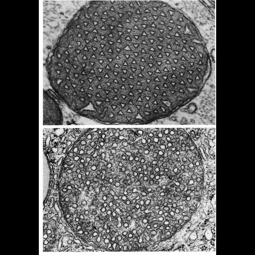
Tubules within the cristae of mitochondria can sometimes take on unusual configurations. Here, in the upper panel, a mitochondrion from an astrocyte in hamster brain appears to have prismatic tubules that are triangular in cross section. The lower panel shows a mitochondrion from rat adrenal cortex with what appears to be vesicular cristae. Arrows point to evidence suggesting that instead, these are probalby tubules with alveolar branches. Description here. Figure 245 (upper) by K. Blinzinger, J. Cell Biol. 25: 293, 1965; fig. 246 (lower) courtesy of Daniel Friend, from Chapter 7 (Mitochondria) of 'The Cell, 2nd Ed.' by Don W. Fawcett M.D. A PDF copy of the accompanying chapter is available on the ASCB's BioEDUCATE website.

Biological Sources

NCBI Organism Classification
Phodopus
Rattus
(Old World rats)
Cell Type
astrocyte
cortical cell of adrenal gland
Cellular Component
mitochondrion
mitochondrial crista
mitochondrial outer membrane
mitochondrial inner membrane

Biological Context

Biological Process
cellular respiration

Attribution

Names
K. Blinzinger
Daniel Friend
Published
ISBN 0721635849
Links
Fawcett's "The Cell" 2nd Ed.
JCB article link
Pubmed
14287181

Grouping

This image is part of a group.
Data Qualifiers
processed data

Sample Preparation

Methods
crosslinking-fixative fixed tissue
embedded tissue
Relation To Intact Cell
microtome-sectioned tissue

Dimensions

Spatial Axis Image Size Pixel Size
X 920px ——
Y 1324px ——
*CIL – Cell Image Library accession number. Please use this to reference an image.